ADVERTISEMENTS:
In this article we will discuss about the anatomy of equisetum. Also learn about its spore-producing organs.
Cut thin transverse sections of different plant parts, stain in safranin-fast green combination, mount in glycerine and observe the anatomical details:
T. S. Internode of Aerial Sterile Shoot:
ADVERTISEMENTS:
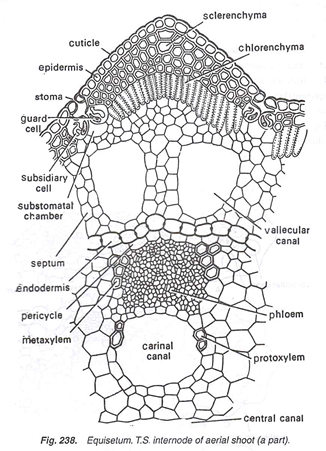
In a cross-section following structures are visible (Figs. 237,238):
1. It is wavy in outline because of the presence of ridges and grooves.
ADVERTISEMENTS:
2. Outermost layer is the epidermis, cells of which have a deposit of silica in their outer and lateral walls.
3. Due to the presence of silica, the stem appears hard and rough to touch.
4. The continuity of epidermis is broken by sunken stomata present in each groove. In each sunken stoma, the guard cells are covered completely by subsidiary cells, thus giving the appearance of two sets of guard cells.
5. Below the epidermis is present a-well-developed cortex.
6. Just below each ridge is present a large patch of sclerenchyma, which is mechanical in function. Sclerenchyma is also present below the grooves in between chlorenchyma (Fig. 237).
7. Inner to be sclerenchyma is present chloren-chymatous tissue below each ridge. It is photosynthetic in function. It extends up to the epidermis in each groove, where lie the stomata.
8. Rest of the cortex is parenchymatous and many layered.
9. Just below each groove is present a large air canal in the parenchymatous cortex. It is known as vallecular canal.
10. Innermost layer of cortex is the endodermis, the cells of which contains casparian strips. But in species like E. sylvaticum, a layer of inner endodermis is also present (Fig. 239 C, D). In E. litorale, each vascular bundle contains its individual endodermis (Fig. 239 E, F).
11. Below the endodermis is present a single-layered pericycle.
12. Vascular bundles are present below the ridges, i.e., alternate to the vallecular canals of the cortex. They are present in the ring.
13. The number of vascular bundles and vallecular canals is equal to the number of ridges and grooves, respectively (Fig. 237).
14. Stele is of ectophloic siphonostelic type.
ADVERTISEMENTS:
15. Each vascular bundle is conjoint, collateral, closed, and consists of xylem, phloem and some parenchyma.
16. In each Vascular bundle is present a water- containing cavity or canal called carinal canal (Fig. 238).
17. Xylem is ‘V’- shaped.
18. Protoxylem is endarch lying opposite to carinal cavity. It consists of annular and spiral tracheids.
ADVERTISEMENTS:
19. Two strands of metaxylem are present.
20. Phloem is present in between two strands of metaxylem and made up of phloem parenchyma and sieve tubes.
21. Pith is present in the form of pith cavity, located in the centre of the aerial shoot (Figs. 237,238).
T.S. Internode of Aerial Fertile Shoot:
ADVERTISEMENTS:
The structure is exactly similar with that of aerial sterile shoot, except a few following minor differences:
1. Absence of stomata.
2. Less developed chlorenchyma and sclerenchyma regions.
T.S. Internode of Rhizome:
It also resembles in structure with the aerial stertile shoot except a few following dissimilarities:
1. Ridges and grooves are not so much well-marked as in sterile shoot.
ADVERTISEMENTS:
2. Absence of stomata (Fig. 240).
3. Absence of chlorenchymatous region.
4. Sclerenchyma is poorly developed.
5. Hollow pith cavity is not well-developed and sometimes it becomes solid.
T. S. Node of Aerial Sterile Shoot:
ADVERTISEMENTS:
It also resembles the internode of aerial sterile shoot except following differences:
1. Absence of all three types of canal, i.e., vallecular canal, carinal canal and central canal.
2. Instead of a central pith cavity, a nodal diaphragm is present (Fig. 241).
3. Vascular bundles are present in the form of a ring after getting fused with each other.
4. Leaf traces and branch traces arise below the ridges and grooves, respectively.
T. S. Adventitious Root:
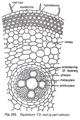
1. Outermost layer is epidermis, from which arise many root hairs.
2. Cortex is thick and multi-layered.
3. Outer zone of cortex consists of 3 to 4-celled thick exodermis.
4. Inner zone is parenchymatous with many intercellular spaces.
5. Endodermis is two-layered (Fig. 242).
6. Pericycle is absent.
7. Stele is a protostele, which is triarch or tetrarch.
8. In the centre is present a large metaxylem tracheid having many protoxylem groups towards the periphery?
9. Phloem is present in between the angles of protoxylem.
Spore-Producing Organs in Equisetum:
Study the external features of cone, cut its transverse and longitudinal sections, stain in safranin, mount in glycerine and study. Also prepare slides of spores and elaters:
Cone:
1. Fertile aerial, unbranched shoots bear at their apices (Fig. 243) the spore-bearing compact organs known as strobili (sing, strobilus) or cones. In some rare cases branched fertile axis is also present (Fig. 244).
2. Each cone or strobilus has a thick central axix known as strobilus axis or cone axis (Figs. 245, 246).
3. On the strobilus axis are attached many umbrella like sporangiophores in whorl.
4. Each sporangiophore is a stalked structure, the free end of which becomes flattened to form a peltate disc (Fig. 245).
5. The disc is a hexagonal structure and present at right angle to the stalk.
6. On the undersurface of disc are present many sporangia, horizontally towards the axis of strobilus.
7. Each sporangium is elongated and pendant, and contains a rounded apex (Fig. 247).
8. The sporangium is surrounded by jacket and tapetal layers which enclose many spores. Equisetum is homosporous (Fig.247).
9. A ring-like outgrowth is present below each strobilus. This is called annulus. Morphological nature of annulus is controversial. According to some it is the uppermost whorl of sterile leaves while others are of the opinion that it is the lowermost sterile sporangiophore (Fig. 246).


