ADVERTISEMENTS:
Study Notes on Cycadopsida. After reading this article you will learn about: 1. Introduction to Cycadopsida 2. Geographical Distribution 3. Characteristics 4. Systematic Position 5. Distribution 6. Identification of Common Indian Species 7. External Morphology 8. Internal Morphology 9. Secondary thickening 10. Leaflets 11. Reproduction 12. Development of Embryo and Other Details.
Introduction to Cycadopsida:
Class-Cycadopsida; Order-Cycadales; Genus-Cycas:
The Cycadales comprise a group of the plants with a number of primitive characters than are possessed by any other group of living gymnosperms, and because of the retention of these primitive characters sometimes they are supposed to be ‘the living fossils’.
ADVERTISEMENTS:
They are of great importance because they occupy the position intermediate between higher cryptogams on one hand and the angiosperms on the other.
The Pteridosperms and the Cycadeoideales are now extinct. It is thought that the Cycadales, were probably derived from the Pteridospermae. The Cycadales is an isolated group among the gymnosperms and in the Cretaceous this group was represented by larger number of species than today.
Geographical Distribution of Cycadopsida:
The Cycadales comprise a single family Cycadaceae which includes nine genera and seventy species. The more interesting point as regards genera is that with the exception of two genera, i.e., Zamia and Cycas, the rest of the genera are very much restricted in their distribution.
ADVERTISEMENTS:
The genera are found in tropical and sub-tropical regions of the world. The four genera, i.e., Zamia, Microcycas, Dioon and Ceratozamia are found exclusively in Western Hemisphere, whereas the remaining five, i.e., Cycas, Macrozamia, Bowenia, Encephalartos and Stangeria in Eastern Hemisphere.
Zamia is found in both northern and southern parts of Western Hemisphere, and the remaining three genera, i.e., Microcycas, Dioon and Ceratozamia are confined in other parts of Western Hemisphere. This way, Zamia is found in both North and South America whereas the remaining three genera grow in localized places of Mexico and West-Indies.
Zamia includes 28 species; Microcycas one species; Dioon three species and Ceratozamia two species.
Cycas, Macrozamia, Bowenia, Encephalartos and Stangeria are confined in Eastern Hemisphere. The genus Cycas is widely distributed and found both in Northern and Southern Hemispheres. They are extended in east from Madagascar to Japan including Australia.
The species of Macrozamia are found only in Australia. Encephalartos and Stangeria are found only in South Africa. Cycas is widely distributed whereas the remaining genera are localized.
ADVERTISEMENTS:
Cycas includes 16 species; Macrozamia 14 species; Bowenia one species; Encephalartos 12 species and Stangeria one species.
Characteristics of Cycadopsida:
1. All cycads are typical xerophytes.
2. The plants are low and palm-like in habit.
3. The stem is short, un-branched, columnar and covered with dense persistent leaf bases.
ADVERTISEMENTS:
4. The leaves are pinnately compound and arranged in a terminal crown.
5. The plants grow very slowly but they live for ages.
6. Comparatively the pith is large and cortex is broad.
7. There is a narrow zone of conducting tissue whereas in conifers the case is reverse. The conducting strand is represented by conjoint, collateral, endarch and open vascular bundles around the pith separated from each other by medullary rays.
ADVERTISEMENTS:
8. The cycads are strictly dioecious, i.e., micro and megasporophylls develop on separate plants. Except the female strobilus of Cycas the sporophylls are arranged in definite cones.
9. The ovules are straight and usually sessile. The pollen chamber is found for receiving the pollens.
10. Male gametes are motile.
The life history of genus Cycas from which the group derives its name is being discussed here.
Systematic Position of Cycas:
ADVERTISEMENTS:
Gymnospermae
Division: Cycadophyta
Class: Cycadopsida
Order: Cycadales
Genus: Cycas
Distribution of Cycas:
Several species of the genus Cycas have been found widely distributed from Madagascar to Japan including Australia. The genus includes about sixteen species which are found wild or cultivated in the tropical and sub-tropical regions of the world.
ADVERTISEMENTS:
In our country they are distributed in the Andaman and Nicobar Islands, Tamil Nadu, Nepal, Sikkim, Bengal and Assam and South Tenasserim. About five species have been reported from various parts of our country.
The species found in our country are:
Cycas revoluta, C. beddomei, C. circinnalis, C. rumphii and C. pectinata.
In general, the plants are low and palm-like. The normal size of the plant ranges from 4 to 8 feet in height. Cycas media is the tallest species upto 20 feet in height. The stem is un-branched, columnar and covered with persistent leaf bases. The leaves are pinnately compound and aggregated in internal crown.
The distinguishing characters of the genus Cycas are as follows:
1. The leaf segment remains circinately involute within the bud.
ADVERTISEMENTS:
2. The pinnae are provided with central mid-rib but no lateral veins.
3. The megasporophylls are not aggregated in cones but borne separately like foliage leaves pectinate in their upper part.
4. The megasporophyll bears on its lower margins two or more ovules.
5. The ovules are ascending.
Identification of Common Indian Species of Cycas:
1. Cycas Revoluta Thunb:
This is native of China and Japan known as sago palm. In India, it is cultivated in the gardens. A palm-like dioecious small tree. Trunk about 1.8 m. to 3 m. high. Suckers present; the stem clothed with old persistent leaf bases. Leaves pinnate, 6 to 1.8 m. long; petiole thick quadrangular; leaflets numerous, sub-opposite, curved downwards, narrow, stiff, dark green, margin revolute, terminating in a spine-like tip.
Carpophylls 10 to 23 cm. long, blade ovate, lacinate into villous segments nearly to the centre, wooly, stalk longer than the blade with 4 to 6 ovules. It flowers in rainy season.
2. Cycas Circinnalis Linn:
This species is commonly found in South India, also cultivated in the gardens. Evergreen palm-like tree upto 8 m. high, cylindrical trunk with a crown of fernlike curved pinnate leaves clothed with prominent annular leaf scars.
Leaves pinnate 1.5 m. to 2.5 m. long; petiole .4 m to .6 m long; leaflets 80 to 100 pairs, alternate, 15 cm to 30 cm. long, margins revolute; carpophylls 18 to 33 cm, x 2.5 to 4 cm.; blade margins triangular with sharp narrow teeth; stalk long bearing in its upper portion 5-18 ovules; male cone about 33 cm long. It flowers from February to March.
3. Cycas Rumphii Miq:
It is found in Andaman and Nicobar Islands, also found in Myanmar, Malayasia, and N. Australia. Evergreen palm-like tree, 3 m. to 7.6 m. high trunk very tough, often branched.
Leaves dark-green and glossy, .6m to .8m leaflets 50-60 pairs, 20 cm. to 38 cm. long, 1.2 cm. to 1.7cm. broad, petiole at base about 4cm. broad; carpophylls about 23 cm. long x 1.2 cm. broad, blade densely villous, ovate male cone 30.4 cm. long and 10 cm. broad; ovules 6 to 10 on the upper portion of the stalk, 10 cm. to 15.2 cm. Flowers in winter season.
4. Cycas Beddomei Dyer:
A small shrub found only in the dry hills of Cuddapah District. A low shrub, the stem 15 cm. high, clothed with leaf bases. Leaves .9 m. long, 23 cm. wide, petiole sub- quadrangular, 15 cm. long leaflets 10 cm. to 17.7 cm. x 3 mm., margins revolute; male cone 30 cm. long with the blade 7.6 cm x 2.5 cm. ovate-lanceolate, dentate bearing two ovules on either side above the middle. Flowering November – December.
5. Cycas Pectinata Griff:
The species is found in the Someshwar hills of Bihar. It is also found in the hills of Assam. It is distributed in Sikkim, Nepal and Pakistan. It is evergreen palm like, 2.4 m. to 3 m. high tree. In Assam, however it attains a height of 6.7 m. Sometimes branched. Leaves 1.5 m. to 2.1 m. long, petiole about 45 cm. long with spinas; leaflets 17.5 cm. to 25.5 cm. long. Male cone about 45.5 cm. x 15.2 cm.
Carpophylls 15.2 cm. long tawny silky, 2 or 3 pairs of ovules above the midrib. Flowers in May.
External Morphology of Cycas:
The Cycas is an Eastern genus. Cycas revolute is found in South India and some parts of Gujarat in natural conditions. This genus differs from other members of Cycadaceae that here female organs do not assemble in a cone while in all other genera the female structure is represented by a simple cone. In general the Cycas plant is un-branched and bearing crown of large fern- like leaves.
The stem of all the species is tuberous when young, but in some species the tuberous body passes into the columnar stem. The stem is usually un-branched but sometimes the branches are seen. The branches are either the result of injury to the stem or it may be the development of the new bud.
The branching may also be the result of the germination of the seeds within the crown of leaves. These new plants send their root into the parent plant body and so the stem appears to be branched.
The Cycas plant grows very slowly, but lives for ages. Usually a new crown of leaves appears at an interval of two to three years, and each such crown remains on the stem for a number of years. When the leaves fall off their bases remain on the stem in the form of a protective armour of leaf-bases.
The leaves of C. revoluta. C media and C. circinnalis are more or less hairy in the bud. The leaves are pinnately compound and like the fern-leaves possess circinate ptyxis. There may be 80 to 100 pairs of leaflets. The fern-like habit of Cycas is noticeable. The bulbils develop in the crevices of the scales which when detached help in vegetative propagation. The bulbils possess a few scales and foliage leaves.
The primary root of Cycas continues as a strong tap root with less laterals. Besides the numerous small secondary soft negatively geotropic coralloid roots develop. These roots are coral-like in appearance and, therefore known as coralloid roots. These roots occur abundantly below the surface of the soil and protruding above it.
Reinke described them as special organs for aeration. Schneider suggested a symbiotic association.
Schaede (1944) investigated the coralloid roots of various cycads and their symbiosis with blue-green algae. It was found that in the species investigated, the coralloid roots whether inhabited by Cyanophyceae or not, contain no bacteria as formerly suggested by Bottomley and Life and other workers.
Schaede (1944) suggested that the bacteria observed by previous workers were probably introduced from the outside by an imperfect technique.
Internal Morphology of Cycas:
Normal Root:
The anatomy of normal root is very simple, the root resembles to that of a dicotyledonous plant in its internal structure. The outermost layer of the roots is known as piliferous layer possessing numerous unicellular root hairs. The root hairs perish soon.
The roots are diarch, triarch or tetrarch. The bundles are radially arranged and the xylem is exarch. In young roots the pith is present but it soon disappears or becomes smaller as soon as the secondary xylem develops due to secondary growth. The secondary xylem is traversed by many parenchymatous rays. Outside the roots the cork is developed.
The cork cambium develops from the pericyclic region.
Coralloid Root:
The anatomy of the coralloid root is similar to that of normal root.
The differences are as follows:
In coralloid root the cortex is divided into three zones, i.e., the outer cortex, middle cortex and the inner cortex. In the outermost region of the root the cork is developed by cork cambium. The outer and inner cortical regions are comprised of parenchyma. The middle cortical zone harbours the blue-green alga Anahaena cycadaeae and is termed algal zone.
The stele is diarch, triarch or tetrarch and surrounded by an endodermis which is followed by pericycle.
Schneider (1894) suggested a symbiotic association of alga and bacteria in the coralloid root. Life (1901) isolated from the roots an alga and some bacteria and believed them to be concerned with the assimilation of nitrogen as well as aeration. Bottomley could isolate nitrogen fixing bacteria from the nodules of Cycas roots.
Stem:
The anatomy of the stem of Cycas resembles to the stem of dicotyledonous angiosperms. The most characteristic feature which distinguishes, the stem of Cycas from other gymnosperms is the presence of a large pith, broad cortex and a narrow zone of conducting tissue.
The cortex is thickly clothed on the outside by the numerous leaf bases and woody scales. The systems of mucilage canals and leaf traces are found in the cortex. The leaf traces are known as the ‘girdles’. In Cycas the leaf traces are concentric and mearch in structure.
The mucilaginous canals are also found in the pith region. The canals of pith are joined to those of cortex by canals running through the medullary rays of the stem.
In central pith region there are many open, endarch vascular bundles separated from each other by medullary rays. The amount of xylem is quite less in the primary stem. The collateral and cauline medullary bundles are found distributed from the pith portion of the stem. At intervals these medullary bundles fuse with each other and form a dense network of anastamosing strands running in pith portion.
Secondary thickening in Cycas:
In Cycas the first cambial ring remains active for a few years and cuts on xylem and phloem in usual way, i.e., xylem towards inside and phloem towards outside. After sometime outside the secondary phloem ring, however, another cambial ring arises at irregular interval.
The first cambial ring by this time ceases to function. This process of secondary growth is repeated at irregular intervals. This way, during the life of Cycas plant a number of alternating rings of xylem and phloem are formed.
The first formed few rings are complete whereas in later stages the cambium begins to cut off only separate vascular bundles. The so formed vascular bundles in later stages by cambium are usually concentric and mesarch. In the case of Cycas the secondary growth does not take place annually and so the rings may not be called the annual rings but they are known as growth rings.
It is also observed that the cambium begins to function when a new crown of leaves is developed, in rest of the period the cambium remains inactive.
The leaf traces curving about through the cortex are known as girdles. Each leaf receives two bundles from the stem stele. A leaf gap is produced whenever a leaf-trace is given off The traces are four in number, two of which are direct and the remaining two pass round the stem through the cortex and often enter a leaf on the opposite side of the stem.
The girdles develop in later stages when the plant becomes old. The leaf traces are concentric and endarch when they start, and mesarch when they pass out the cortex.
The presence of leaf gaps and concentric bundles denotes prominent feature of pteridophytes, which shows a strong affinity between Cycas and ferns.
Rachis:
The rachis of the leaf of Cycas is quite stout and woody. To study the internal structure of the rachis the transverse sections are required. The outermost layer is the single layered epidermis covered by a thick cuticle. Just beneath the epidermis there is a hypodermis which is two or three layers in thickness on the adaxial side and many layered on the abaxial surface.
The cells of the hypodermis are sclerenchymatous. The ground tissue which follows the hypodermis is parenchymatous.
The most characteristic feature of the rachis is the arrangement of vascular bundles in a omega-shaped (Ω) outline. The vascular bundles vary in their structure at the base, centre and apex of the rachis. Each vascular bundle is ensheathed within a single layered sclerenchymatous sheath.
The vascular bundles are open and collateral. A single layered endodermis and one or multi-layered pericycle surround the vascular bundle.
The interesting changes take place at the base of the rachis when the transition from the stem type of xylem takes place into the petiolar arrangement. The bundles are endarch at the base, i.e., the protoxylem remains towards the centre. Here, the xylem develops centrifugally. The phloem lies towards the abaxial side. The cambium is found at the upper surface of the centrifugally developed metaxylem.
During the upward course of the bundles in the rachis the metaxylem tracheids appear opposite the protoxylem and constitute the centripetally formed metaxylem. Here, the metaxylem is present on both the sides of protoxylem and the bundles are mesarch.
Simultaneously, with the appearance of the centripetal metaxylem elements the cambium begins to function on the inner side. In further upward course the centripetally developed metaxylem increases in quantity and on the other hand the centrifugal metaxylem decreases and only two patches of them remain, each consisting of a few tracheids only.
All along the phloem remains towards abaxial side. This way from the base to the apex of the rachis the bundles change their position of xylem elements from endarch to exarch.
Leaflets of Cycas:
The leaflet of the Cycas leaf is leathery and thickly cutinized. It possesses all xerophytic characters. The epidermis and the upper hypodermis are highly thickened. On the lower surface the sunken stomata are situated in pits with overarching rims. Just beneath the hypodermis there is mesophyll tissue.
The mesophyll has a well-developed palisade layer on its upper side. The lower part of the mesophyll consists of parenchymatous cells with scanty intercellular spaces.
In between paliside layer and lower mesophyll there is a three or four celled thick layer of transversely running long colourless cells from the mid-rib to near the margin. These cells constitute the transfusion tissue. In the case of Cycas the mid rib is un-branched and there are no lateral veins and therefore, the transfusion tissue form a conducting channel for water, and thus the name transfusion tissue is derived.
The spongy tissue is found below the transfusion tissue. This helps in aeration. The central portion of the leaflet is bulged which indicates the position of the mid-rib. The vascular tissues are confined to the mid-rib only. The mid-rib bundle consists of a broad triangular centripetally developed exarch xylem and two small patches of centrifugally developed endarch primary xylem groups.
They are known as centripetal and centrifugal xylem patches. The presence of the diploxylic structure is the characteristic feature of this genus. The phloem is found on abaxial side.
The pericycle is parenchymatous. The endodermis is single layered and sclerenchymatous which ensheaths the bundle. In between phloem and centripetal xylem there lies a cambium. The phloem consists of sieve tubes and phloem parenchyma, the companion cells are altogether absent. Calcium oxalate crystals are found in various cells of the mid-rib.
The remaining part of the bundle is occupied by parenchyma. A number of tracheidal cells are found on either side of the vascular bundle.
Reproduction of Cycas:
The reproduction in Cycas takes place by means of vegetative and sexual methods.
Vegetative Reproduction:
The vegetative reproduction takes place by means of bulbils which develop in the crevices of the scales, when detached help in vegetative propagation. Each bulbil possesses a few scales and foliage leaves.
Sexual Reproduction:
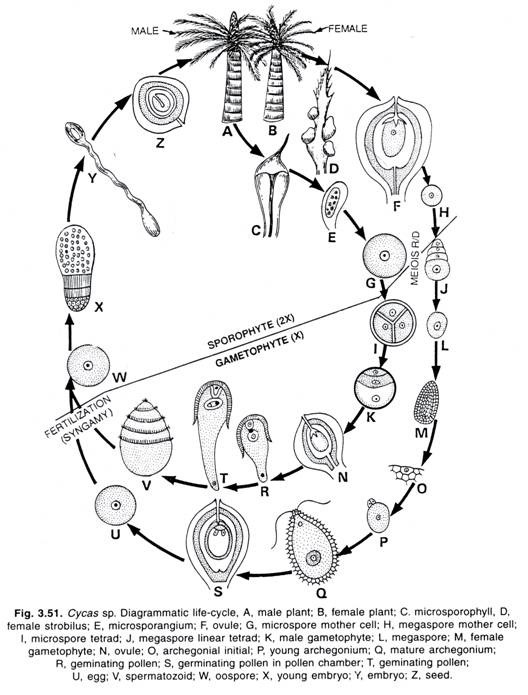
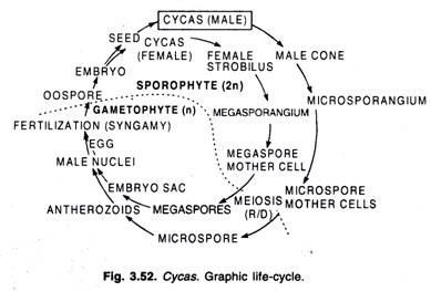
The Cycas is strictly dioecious plant, i.e., male and female organs are found on two separate plants. The microspores (male) and megaspores (female) develop in micro and mega-sporangia respectively. The male plants of Cycas are very rare in our country. The cones or strobili are found at the apex of the trunk concealed at first among the bases of the leaves.
The male flowers are found to be arranged in erect cones. Usually a single male cone develops at the apex of male plant. The megasporophylls or female strobili are found in crowded whorls around the apex of the female plant alternating with whorls of foliage leaves.
Male Cone:
The male cone is about more than 50 cm. in length. Usually a single male cone develops at the apex of the plant. The sporophylls are closely imbricate and spirally arranged in a compact cone. Some of the sporophylls of the base and apex of the cone are being sterile.
Each microsporophyll is narrow below and broad above terminating into a projection, the apophysis. The microsporangia are confined to the abaxial (lower) surface of the microsporophyll, the upper surface is sterile. The sporangia may number upto one thousand in certain species.
The sporangia occur on two flanks of the microsporophyll differentiated by a median sterile ridge. They are usually found in definite sori each consisting of two to six sporangia. The sporangia are more or less united at the base. The microsporangia develop from the hypodermal cells.
The wall of sporangium is four to seven layered. There is definite tapetal layer developed from the sporogenous tissue for the nourishment of the microspores. The sori of microsporangia are intermingled with hair.
The last generation of the sporogenous tissue forms the spore mother cells. Each spore mother cell divides meiotically and four microspores or pollens are produced. A mature microspore possesses two sharply distinguished coats known as exine and intine. A large number of microspores are produced in each sporangium.
The microspore germinates in situ, i.e., within the microsporangium. At the time of the pollination the nuclear division has already taken place.
Female Strobilus:
In Cycas the female strobilus is made up of a whorl of spirally arranged megasporophylls which are situated among the crown of vegetative leaves. The megasporophylls are not arranged in definite cones. The megasporophyll of Cycas revoluta is one of Nature’s curiosities.
It preserves in a living seed plant the image of the seed-bearing fronds of Palaeozoic pteridosperms. The megasporophylls of Cycas resemble to the foliage leaves.’
The apical meristem after producing megasporophylls produces vegetative leaves. The crown of megasporophylls develops each year in between successive crown of foliage leaves. The megasporophylls develop only when the plant is quite old. The female strobilus consists of a rosette of sporophylls resembling reduced foliage leaves. Here, the ovules replace the lower pinnae or spines.
The megasporophylls of Cycas revoluta resemble too much to the vegetative leaves than in any other species of Cycas. In C. circinnalis, more reduction taken place but the pinnate character of vegetative leaf still persists. In Cycas revoluta and C. circinnalis several ovules develop on the margins of the megasporophyll whereas in C. siamensis only two ovules are found.
In C. rumphii the magasporophyll bears two to five ovules. The ovules of Cycas are quite large. In Cycas circinnalis, the largest ovules are formed. Each ovule is 6 cm. X 4 cm. The ovules of C. circinnalis are smooth and dark-green, whereas of C. revoluta are densely hairy and orange-red in colour.
The Ovule:
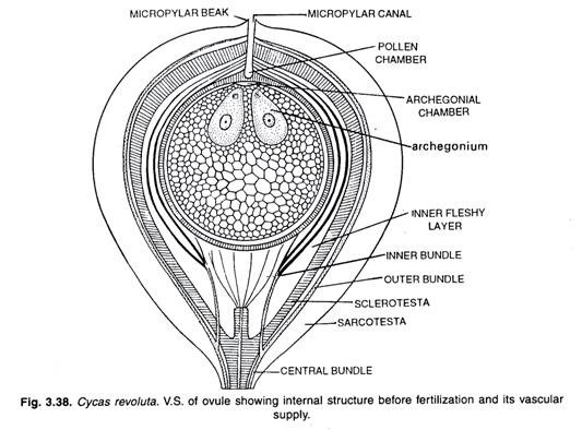
The ovules are sessile and orthotropous in their position. They are ovoid or spherical and vary in their size. The ovule of Cycas circinnalis may attain the length of 6 cm. Each ovule has a single thick integument which is differentiated in three layers. The inner and outer fleshy layers of the integument remain separated from each other by a hard stony layer in between the two.
The inner fleshy layer remains more prominent in earlier stages of the development of the ovule. On the ripening of seed it more or less disappears.
There is a mass of nucellar tissue of nucellus within the integument. The nucellus remains fused with the inner fleshy layer of the integument except at the tip portion. In the top portion of the ovule the integument forms a long micropyle. In the apical portion the nucellus forms a nucellar beak.
The beak forces its way into the micropyle, while in the centre of the beak and below it, cells break down and form the pollen chamber.
The ovule is well supplied with vascular bundles. The bundles serve the base of the ovule. Each vascular bundle divides into two branches, each serving the outer and inner fleshy layers. These branches further divide for proper vascular supply.
The ovule arises as a hypodermal mass of meristematic cells. The cells above it divide rapidly and form the nucellus. The surrounding cells also divide rapidly and the nucellus becomes embedded within these cells. A thick integument develops outside the nucellus. A single large megaspore develops deep within the nucellus.
Megaspore:
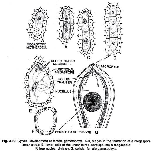
In the beginning all the cells of nucellus are quite-alike. They are thin-walled and parenchymatous. Soon after, one of the cells in the central region of the nucellus becomes prominent. This bigger prominent cell acts as megaspore mother cell. The megaspore mother cell divides meiotically and a linear tetrad of four cells is resulted.
This way the megaspore mother cell represents the last stage of sporophytic generation. The three upper cells or megaspores of the linear tetrad degenerate and the basal functional megaspore increases in size. The upper three degenerating megaspores are nutritional in function. They provide the nutrition for the development of the functional megaspore. The megaspore is also known as embryo-sac.
Development of Female Gametophyte:
The megaspore increases in size and gradually continues to absorb some of the neighbouring cells of the nucellus. The surrounding cells of the nucellus form a spongy nutritive tissue around the developing megaspore. Free nuclear division takes place until a thousand free nuclei are formed within the megaspore.
A large central vacuole appears and the nuclei are now being pushed towards periphery. After the peripheral shifting of the nuclei, the wall formation begins to take-place. The wall formation begins at the periphery of the embryo sac and advances towards the centre. Ultimately whole of the megaspore of embryo sac fills up with cellular tissue which in earlier stages contain sugar and in later stages starch.
Some cells of the female gametophyte contain tannin in them. While the megaspore increases in size, its germination starts. The surrounding nucellar tissue forms a layer around the germinating megaspore which is nutritive in function and known as endosperm jacket.
This layer closely resembles to the tapetum of the other plants. The food material from the nucellus passes through this endospermic jacket layer for the development of the germinating megaspore.
The archegonia begin to develop on the germinated megaspore. Usually the number of archegonia is lesser than ten and they are confined to micropylar end of female gametophyte. They are usually three to five in number. The archegonial cells are however, more in number.
Development of Archegonium:
The superficial archegonial initial divides by a transverse wall resulting into a primary neck cell and a central cell. The primary neck cell divides vertically forming two neck cells. These neck cells form a small neck. The central cell enlarges in size and becomes vacuolated. Soon after this gets surrounded by an archegonial jacket which acts as nutritive layer.
The pits with fine strands of protoplasm connecting the jacket cells and the egg have been observed in Cycas circinnalis, and large quantity of food passing through these pits into the central cells has also been observed. The central cell divides into two cells known as ventral canal cell and the egg. Soon after, the ventral canal cell disappears.
Towards the micropylar end, just at the base of micropyle a small depression appears at the place where the archegonia are developed. The surrounding endospermic tissue is gradually absorbed and the mature archegonia open into a cavity, the archegonial chamber.
The over-lined nucellar tissue has already been absorbed; the megaspore membrance has already been ruptured and the archegonia open into the archegonial chamber.
Pollination and Development of Male Gametophyte:
The Cycas is wind pollinated. At the time of pollination, the nuclear division of the microspore results in the formation of a small persistent prothallial cell and a large antheridium initial. Soon after the nucleus of the antheridium initial divides into two unequal cells. The small cell which remains in close association of the prothallial cell is known as generative cell, whereas the other bigger cell is the tube-cell.
This way, the microspores are three-celled at the time of pollination. At this stage some of the nucellus cells degenerate forming a pollen chamber. A mucilaginous pollination drop oozes out from the micropyle. In the liquid, a large number of pollen grains (microspores) are caught and, as it dries up they are drawn into the pollen chamber. Later on the apex of nucellar beak becomes very hard.
About a week after pollination, the pollen germinates in the pollen chamber. The tube cell increases in size and the exine of the pollen grain bursts giving the way to the inline in the form of a pollen tube. The haustorial end penetrates the nucellus.
The haustorial end is the end of the pollen from which the pollen tube is given off In Cycas revoluta, the time in between pollination and fertilization under natural conditions is of about four months.
The generative cell divides into the stalk cell and the body cell. The stalk cell does not divide further, but increases in size and is full of starch grains. The stalk cell is sterile cell and regarded as representing the actual stalk cell of an antheridium.
The body cell enlarges sufficiently; however, this does not contain starch grains. Two blepharoplasts appear at the two poles of the nucleus of the body cell. The blepharoplasts are small, star-shaped and protoplasmic bodies.
They appear a few months before the division of the body cell. The blepharoplasts are concerned in the production of cilia; they become vacuolated and break up into a mass of granules which fuse and finally give rise to five or six spiral bands along with numerous cilia are arranged.
In the earlier stages the blepharoplasts lie in the plane of long axis, but later on with the enlargement of the pollen tube and body cell they rotate through 90° and come to lie transverse to the long axis of the pollen tube.
Ultimately the body cell divides into two forming two antherozoids. The sperms of Cycas are the largest among plants. They are easily visible to the naked eye. The movement of the sperms begins even when they are still within the mother cells.
The sperms are liberated and they swim actively within the pollen tube. The pollen tube becomes turgid and ultimately ruptures. Ikeno described sperms of Cycas to be completely naked. According to Swamy (1948) the pollen tube does not burst but simply an aperture is formed at the end of the tube and the spermatozoids are squeezed through it by amoeboid movements.
Fertilization:
On breaking of the pollen chamber through the base of the nucellar cap, the archegonial chamber and the pollen chamber merge together forming a continuous cavity. The archegonial chamber does not contain any liquid but is moist.
On rupturing the pollen tube the liquid is discharged and the sperms swim in this liquid till they enter the necks of archegonia. In earlier stages of the development the pollen tube acts as a haustorium.
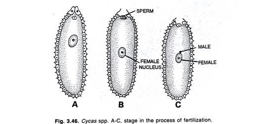
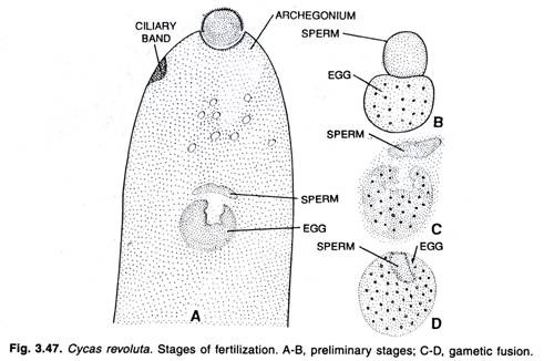
The spermatozoids swim in the liquid discharged from the pollen tubes and enter the necks of archegonia with a sufficient force. The entire sperm enters the egg but soon after the cytoplasmic sheath and ciliated band are removed and the naked male nucleus moves to the large egg nucleus.
The male and female nuclei are united and the fertilization is effected. With the result of union the oospore is formed. The oospore is diploid (2n) and this represents the beginning of the sporophyte generation. The fertilization which is furnished by means of a pollen tube is known as siphonogamy. In Cycas, the sperms are motile and, therefore, pollen tube acts as a haustorium and not as a sperm carrier.
Development of Embryo of Cycas:
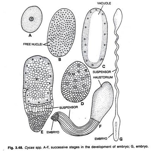
The oospore undergoes free nuclear division like that of germinating megaspore, resulting in the formation of 200 to 300 nuclei distributed throughout the cytoplasm. In Cycas revoluta there are certainly as many as 256 nuclei and very probably more.
According to Treub (1884), in Cycas circinnalis a large central vacuole appears in the centre and all the nuclei are pushed to the periphery so that they form a parietal layer. In Cycas revoluta, Ikeno in 1898 observed the formation of the central vacoule in a different way.
He stated that a large number of small vacuoles are formed, which result in the formation of the central portion of the proembryo containing nuclei and a parietal layer of cytoplasm and nuclei. The central portion of the proembryo with its nuclei disorganizes leaving a parietal layer of cytoplasm and nuclei and a large central vacuole.
According to Ikeno (1898) in Cycas revolute, simultaneously free nuclear divisions at the base of the proembryo take place before the cell walls begin to appear, permanent cell walls are laid at the base of the proembryo and the cells of this region become differentiated into three regions.
The cells encircling the central vacuole become more or less haustorial; the cells immediately beneath get elongated and form suspensor, while the cells at the tip remain meristematic and give rise to the embryo proper.
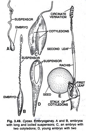
The suspensor is sufficiently long but it does not branch or bear more than one embryo. The suspensors separate from the several archegonia but soon after become twisted together, so that it appears a single suspensor composed of two, three or more suspensors.
The extra suspensors are terminated by abortive embryos; only the successful embryo stretches in suspensor beyond the level of abortive embryos. The single successful embryo rapidly grows into the tissue of endosperm and increases in size for some time. At the base of the embryo there develops a peculiar structure known as coleorhiza and at the opposite end the stem tip and two cotyledons are differentiated.
Seed of Cycas:
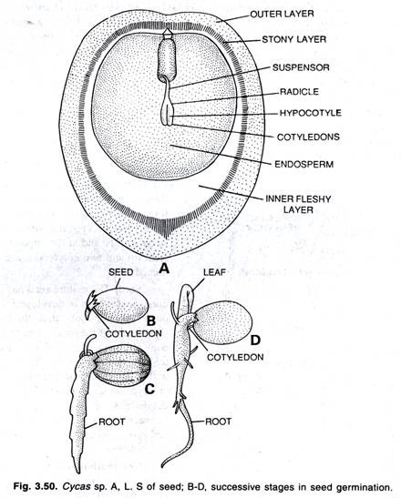
The mature seed has a fleshy outer coat which is developed from the outer portion of the three-layered integument of the ovule. The inner stony coat of seed is developed from the middle stony layer of the integument, within the seed coat there lies a straight embryo, which consists of two unequal cotyledons.
There lies a plumule in between two cotyledons. The axis of the embryo is differentiated into hypocotyl and radicle.
Germination of Seed (Seedling):
The seeds have no resting period and germinate immediately if placed on moist soil. The outer fleshy coat of the seed may persist for months even after the seed has started its germination. The seed is large and longer than broad. It falls on the soil with long axis parallel to the earth. It absorbs water from the moist soil and the coleorhiza comes first and the tip of the primary root makes its way through the coleorhiza.
The major part of the cotyledons comes out of the seed, but the tips of the cotyledons remain within the seed for considerably a long time till the whole of endosperm is transformed on to the cotyledons. The leaves begin to develop singly on the new plants and it has been observed that for a number of years only one leaf is developed at a time.
It is after several years that a crown of leaves begin to develop at the apex of the stem. In the beginning the number of the leaves is quite less and after several years the normal crown of the leaves is formed. In Cycas, the leaves during development show circinate vernation like those of ferns.
Origin and Relationships:
According to Arnold (1953), the modern cycads are the remnant of a much larger vascular plant group that during the past ranged far beyond its present limits. Every genus probably evolved independently, within or near its present general range and none arose from any of the others. The genus Cycas has been considered the most primitive because of the lack of definite ovulate strobili.
The Cycadales are believed to have evolved from pteriodosperms (Cycadofilicales) during the later part of the Carboniferous period, but the particular pteridosperm which served as ancestor has not been identified.
Resembled with other Gymnosperms
Cycadofilicales resembled with the Filicales and Cycadales. On one hand the Cycadofilicales resembled the ferns in their general habit, vascular anatomy and occurrence of sporangia in son while on the other hand they resembled with the cycads in the structure of their ovule.
The ovules in both the cases possess three-layered integument, the two vascular strands traversing the outer and inner fleshy layers and the nucellar beak with a distinct pollen chamber.
The Bennettitales of Mesozoic era had several affinities with Cycadofilicales, ferns and cycads. It seems quite clear that as regards their origin the cycads are connected with the extinct Bennettitales. However, these two gymnospermic groups have so many features in common and therefore, for this reason Bennettitales have been called fossil cycads.
The stem of the Cycadales, like that of the Bennettitales, is an endarch siphonostete with a narrow zone of wood, the xylem in other aerial parts of the plant being mesarch.
The dicotyledonous embryo of Cycadales is also suggestive of that of Bennettitales. The Bennettitales began to come in prominance as the Cycadofilicales declined, with the result Bennettitales carried forward many of the primitive features of the Cycadofilicales.
Cycadales best flourished in Mesozoic. The Cycads resemble the ferns in their general habit, vascular anatomy, structure and venation of the leaves, occurrence of the sporangia in sori, structure of the microsporangia and multi-ciliate sperms. All the above mentioned characters and the structure of ovule, are common to the Cycadofilicales, Bennettitales and Cycadales.
Hence it is thought that three orders are closely related with each other and represent a distinct line of evolution reaching far back into the Palaeozoic. Cycas being the most primitive of living Cycadales, on account of its loose and fern like megasporophylls, stand nearest to the fossil seed plant line and therefore, the Cycas (cycads) is thought to be a ‘living fossil’.
Economic Importance of Cycas:
The pith of Cycas revoluta yields sago and the fruits can be eaten, being rich in protein and soluble non-nitrogenous substances. In Europe, the leaves of Cycas revoluta after silvering and treatment in various ways are made into funeral wreaths, there the leaves are called ‘palm leaves’. The leaves of C. circinnalis are used in making mats in South India.
The young shoots of C. circinnalis are eaten, and sago is also obtained from the trunk.
For obtaining the sago, the trunk is cut into disks which when dry are pounded into flour and thereafter thrown into water where the starch settles down. The large fruits of C. circinnalis yield annually about the same quantity of starch. The hill tribes of Assam eat the seeds and tender fleshy shoots of Cycas pectinata.
The fleshy stem of C. pectinata is pounded and used as a hair wash for diseased root hair. The sago is also extracted from the trunk of Cycas rumphii. The cooked fruits of C. rumphii are eaten by Andamanese tribes while the uncooked fruits are poisonous.


