ADVERTISEMENTS:
Let us learn about Cambium. After reading this article you will learn about: 1. Origin of Cambium 2. Fascicular and Inter-fascicular Cambium 3. Duration 4. Functions 5. Structure 6. Cell Division 7. Thickening in Palms.
Origin of Cambium:
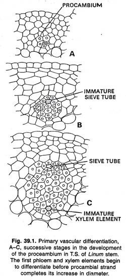
The primary vascular skeleton is built up by the maturing of the cells of the procambium strands to form xylem and phloem. The plants which do not possess secondary growth, all cells of the procambium strands mature and develop into vascular tissue.
In the plant which have secondary growth later on, a part of the procambium strand remains meristematic and gives rise to the cambium proper. In roots the formation of cambium differs from that in stems because of the radial arrangement of the alternating xylem and phloem strands.
ADVERTISEMENTS:
Here the cambium arises as discrete strips of tissue in the procambium strands inside the groups of primary phloem. Later on, the strips of cambium by their lateral extension are joined in the pericycle opposite the rays of primary xylem. The secondary tissue formation is most rapid beneath the groups of phloem so that the cambium, as seen in the transverse section of older roots, soon forms a circle.
Fascicular and Inter-fascicular Cambium:
In stems the first procambium that develops from promeristem is usually found in the form of isolated strands. In some plants these first-formed strands soon become, united laterally by additional similar strands formed between them and by the lateral extention of the first-formed strands.
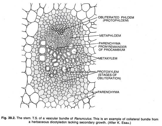
During further development this procambial cylinder gives rise to a cylinder of primary vascular tissue (xylem and phloem) and cambium. Later on, a cylinder of secondary vascular tissue is formed that arises in strands as does the primary cylinder. In Ranunculus and some other herbaceous plants, the procambium strands, and the primary vascular tissues, do not fuse laterally but remain as discrete strands.
ADVERTISEMENTS:
More often in herbaceous stems the cambium extends laterally across the intervening spaces until a complete cylinder is formed. Where such extension occurs, the cambium arises from inter-fascicular meristematic cells derived from the apical meristem.
The strips of cambium that arise within collateral bundles are known as fascicular cambium, and the cambial strips found in between the bundles are known as inter-fascicular cambium.
Duration of Cambium:
The duration of the functional life of the cambium varies greatly in different species and also in different parts of the same plant. In a perennial woody plant the cambium of the main stem lives from the time of its formation until the death of the plant.
It is only by the continued activity of the cambium in producing new xylem and phloem that such plants can maintain their existence. In leaves, inflorescenes and other deciduous parts, the functional life of the cambium is short. Here all the cambium cells mature as vascular tissue. The secondary xylem is directly found upon the secondary phloem in such bundles.
Function of Cambium:
The meristem that forms secondary tissues consists of an uniseriate sheet of initials that form new cells usually on both sides. The cambium forms xylem internally and phloem externally. The tangential division of the cambial cell forms two apparently identical daughter cells.
One of the daughter cells remains meristematic, i.e., the persistent cambial cell, the other becomes a xylem mother cell or a phloem mother cell depending upon its position internal or external to the initial. The cambium cell divides continuously in a similar way; one daughter cell always remains meristematic, the cambium cell, whereas the other becomes either a xylem or a phloem mother cell.
Probably there is no definite alternation and for brief periods only one kind of tissue is formed. Adjacent cambium cells divide at nearly the same time, and the daughter cells belong to the same tissue. This way, the tangential continuity of the cambium is maintained.
Structure of Cambium:
ADVERTISEMENTS:
There are two general conceptions of the cambium as an initiating layer:
1. That it consists of a uniseriate layer of permanent initials with derivatives which may divide a few times and soon become converted into permanent tissue;
2. That there are several rows of initating cells which form a cambium zone, a few individual rows of which persist as cell forming layers for some time. During growing periods the cells mature continuously on both sides of the cambium it becomes quite obvious that only a single layer of cells can have permanent existence as cambium.
Other layers, if present, function only temporarily and become completely transformed into permanent cells. In a strict sense, only the initials constitute the cambium, but frequently the term is used with reference to the cambial zone, because it is difficult to distinguish the initials from their recent derivatives.
Cellular Structure of Cambium:
There are two different types of cambium cells:
1. The ray initials, which are more or less isodiametric and give rise to vascular rays; and
2. The fusiform initials, the elongate tapering cells that divide to form all cells of the vertical system.
ADVERTISEMENTS:
The cambial cells are highly vacuolated, usually with one large vacuole and thin peripheral cytoplasm. The nucleus is large and in the fusiform cells is much elongated. The walls of cambial cells have primary pit fields with plasmodesmata. The radial walls are thicker than tangential walls, and their primary pit fields are deeply depressed.
Cell Division in Cambium:
With the result of tangential (periclinal) divisions of cambium cells the phloem and the xylem are formed. The vascular tissues are formed in two opposite directions, the xylem cells towards the interior of the axis, the phloem cells toward its periphery. The tangential divisions of the cambium initials during the formation of vascular tissues determine the arrangement of cambial derivatives in radial rows.
ADVERTISEMENTS:
Since the division is tangential, the daughter cells that persist as cambium initials increase in radial diameter only. The new cambium initials formed by transverse divisions increase greatly in length; those formed by radial divisions do not increase in length.
As the xylem cylinder increases in thickness by secondary growth, the cambial cylinder also grows in circumference. The main cause of this growth is the increase in the number of cells in tangential direction, followed by a tangential expansion of these cells.
Cambium Growth about Wounds:
One of the important functions of the cambium is the formation of callus or wound tissue, and the healing of the wounds. When wounds occur on plants, a large amount of soft parenchymatous tissue is formed on or below the injured surface; this tissue is known as callus. The callus develops from the cambium and by the division of parenchyma cells in the phloem and the cortex.
During the healing process of a wound the callus is formed. In this there is at first abundant proliferation of the cambium cells, with the production of massive parenchyma. The outer cells of this tissue become suberized, or periderm develops within them, with the result a bark is formed.
However, just beneath this bark the cambium remains active and forms new vascular tissue in the normal way. The new tissue formed in the normal way extends the growing layer over the wound until the two opposite sides meet. The cambium layers then unite and the wound becomes completely covered.
ADVERTISEMENTS:
Cambium in Budding and Grafting:
In the practices of budding and grafting, the cambium of both stock and scion gives rise to callus which unites and develops a continuous cambium layer that gives rise to normal conducting tissue. There is an actual union of the cambium of stock and scion of two plants during the practices of budding and grafting and therefore these practices are not commonly found in monocotyledons.
Cambium in Monocotyledons:
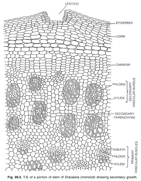
A special type of secondary growth occurs in few monocotyledonous forms, such as Dracaena, Aloe, Yucca, Veratrum and some other genera. In these plants the stem increases in diameter forming a cylinder of new bundles embedded in a tissue.
Here a cambium layer develops from the meristematic parenchyma of the peri-cycle or the innermost cells of the cortex. In the case of roots, the cambium of this develops in the endodermis. The initials of cambium strand in tiers to form a storied cambium as found in the normal cambium of some dicotyledons.
Cambium in Thickening in Palms:
ADVERTISEMENTS:
The palm stems do not increase in girth, because of any cambial activity but this thickening is the result of gradual increase in size of cells and of intercellular spaces and sometimes of the proliferation of fibre tissues. This is the type of long continuing primary growth.
The process is as follows:
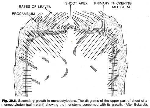
Most of the monocotyledons lack secondary growth, but with the result of intense and long continuing primary growth they may produce such large bodies as those of the palms. The monocotyledons often produce a rapid thickening beneath the apical meristem by means of a peripheral primary thickening meristem as shown in figure.
The activity of the primary thickening meristem resembles with secondary growth found in certain monocotyledons such as Dracaena, Yucca, etc. The apical meristem also known as shoot apex produces only small part of the primary body, i.e., a central column of parenchyma and vascular strands.
Most of the plant body is formed by the primary thickening meristem. The primary thickening meristem is found beneath the leaf-primordia, which divides periclinally producing anticlinal rows of cells. These cells differentiate into a tissue formed of ground parenchyma traversed by procambial strands.
These procambial strands later on develop into vascular bundles. The ground parenchyma cells enlarge and divide repeatedly, causing increase in thickness. This way, both apical meristem and primary thickening meristem give rise to the main bulk of the stem tissues of monocotyledons.
The thickening takes place in monocotyledons, such as palms, due to the activities of the apical meristem and primary thickening meristem.


