ADVERTISEMENTS:
In this article we will discuss about the vegetative and sexual modes of reproduction in Riccia.
Vegetative Reproduction:
Vegetative Reproduction of Riccia takes place by the following methods:
(a) By Progressive Death and Decay of the Older Parts of the Thallus (Fragmentation):
ADVERTISEMENTS:
Vegetative propagation is taking place usually by the continuous growth from the growing point. The older basal parts of dichotomies die and rot away, while the tip grows continuously to give separate plants. The growing tip survives the dry period one season, then grows next year.
(b) By Adventitious Branches:
Vegetative growth of Riccia also takes place by the formation of adventitious branches from the lower surface of the thallus. The branch that separates from the parent thallus, grows into new plant (viz., Riccia fluitans).
(c) By Tubers:
ADVERTISEMENTS:
In many species (viz. R. bulbifera, R. discolor, R. perennis, R. vesicata) tubers develop on the thallus. These perennating structures can easily survive a period of drought and germinate in favourable season.
(d) By Rhizoids:
It is found that new gametophytes develop from the tips of the rhizoids (e.g. R. glauca). The tips of the rhizoids divide repeatedly to form a mass of chloro- phyllous cells. These cells further grow to form new thallus.
Sexual Reproduction:
Sexual reproduction takes place by means of well-developed sex organs: the male reproductive organ, antheridium and the female, archegonium. Riccia are mostly monoecious or homothallic (e.g. R. melanospora, R. crystallina, R. cruciata, R. pathankotensis, R. glauca and R. robusta), but some are also dioecious or heterothallic (R. discolor, R. sanguinea, R. frostii, R. pearsonii).
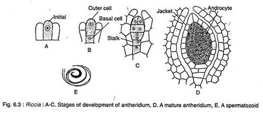
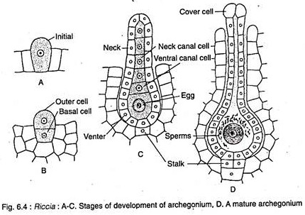
Sex organs develop singly. They are formed acropetally in a linear row on the upper median furrow (Fig. 6.1 B-C). At first they are superficial, but at later stages of development they are enveloped by the outgrowth of tissues and are finally embedded in a cavity formed by the overarching tissues (Fig. 6.3D and 6.4D).
These cavities are called antheridial and archegonial chambers that are open on the upper surface by narrow cylindrical canals.
ADVERTISEMENTS:
Antheridium:
Development and Structure of Antheridium:
The antheridium develops from a single superficial dorsal cell, called the antheridial initial (Fig. 6.3A) which lies 2 or 3 cells behind the apical cell and placed in the median furrow. The antheridial initial divides transversely to form an upper outer cell and a lower basal cell (Fig. 6.3B).
The outer cell later develops the antheridium with jacket and the basal cell forms the stalk (Fig. 6.3C). The tissue surrounding the antheridial initial tends to overgrow and ultimately envelops the antheridium leaving a pore at top.
ADVERTISEMENTS:
The mature antheridium (Fig. 6.3D) is globular or pear-shaped with a single-layered jacket (wall) “enclosing a number of androcyte mother cells. Each androcyte mother cell divides diagonally to form two triangular androcyte cells (Fig. 6.3D).
Each androcyte cell forms a biflagellated spermatozoid or antherozoid (Fig. 6.3E). At maturity all the cell walls inside the antheridium disintegrates. The antherozoids are now free inside the antheridial jacket in a gelatinous fluid.
Archegonium:
ADVERTISEMENTS:
Development and Structure of Archegonium:
Like antheridium, the archegonium also develops from a single superficial dorsal cell, called the archegonial initial (Fig. 6.4A), placed in the median furrow and close to the apical cell. The archegonial initial divides transversely forming an upper outer cell and a lower basal cell. The outer cell later develops into archegonium proper, while the basal cell forms the stalk (Fig. 6.4B & C).
The mature archegonium (Fig. 6.4C) is a flask-shaped structure having a short stalk, a swollen basal venter containing a large egg or ovum, a ventral canal cell and an elongated neck containing a row of four neck canal cells.
The tip of the neck is covered by four specialised cover cells that are larger than the remaining cells of the neck. The archegonium is not fully embedded in the archegonial chamber as in the case of the antheridium, so that the tip of the archegonial neck protrudes out.
ADVERTISEMENTS:
Fertilisation of Archegonium:
At maturity, the neck canal cells and the ventral canal cell of archegonia disintegrate to form a mucilaginous mass. The mucilage imbibes water, swells and comes out by forcing upon the cover cells, forming a narrow passage called the neck canal.
In this condition, fertilisation takes place which requires the presence of water that acts as a medium for swimming the antherozoids. Water is also essential for the disintegration of the antheridial jacket vis-a-vis liberation of antherozoids. After rain or heavy dew, water is usually retained in the dorsal groove of the thallus in the form of thin film.
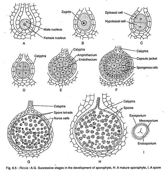
The liberated spermatozoids swim in this water and they come near the archegonium due to chemotactic attraction induced by proteins and other chemicals coming out of hollow archegonial neck (Fig. 6.4D). A number of sperms pass into the neck, reach the venter and ultimately one fertilises the egg (Fig. 6.5A-B) to form a diploid zygote or oospore, thus the sporophytic generation begins.
The Sporophyte:
The zygote invests itself with a thin cellulose wall immediately after fertilisation. It increases greatly in size and almost completely fills up the venter of the archegonium (Fig. 6.5B). Simultaneously, the walls of the venter divide periclinally and eventually form a two-layered calyptra around the young sporophyte.
Development of the Sporophyte:
ADVERTISEMENTS:
The first division of the zygote is transverse (Fig. 6.5C). The second division, at right angles to the first one, results in the formation of a quadrant stage of four cells (Fig. 6.5D). Another division in a vertical plane at right angles to the former resulting in an octant stage of eight cells. Subsequent divisions are in all directions giving rise to a spherical mass of 20 to 30 cells.
The superficial cells of this mass divides periclinally to form an outer amphithecium layer and a inner mass of endothecium (Fig. 6.5E). The amphithecium forms the jacket of the sporophyte, while the endothecium represents the archesporium (Fig. 6.5F).
The archesporial cells divide further to form two types of cells:
(a) The spore mother cells (sporocytes) and
(b) The sterile nurse cells with a watery vacuolated cytoplasm (Fig. 6.5G). These nurse cells are probably the forerunners of elaters.
The spore mother cells or sporocytes are the last cells of the sporophytic generation after which it begins to disintegrate. The nurse cells and the amphithecial jacket layer also disintegrate further followed by disintegration of the inner cell layers of the venter wall (Fig. 6.5H). All the disintegrated products form a nutritive fluid within which the spore mother cells remain suspended.
ADVERTISEMENTS:
Sporogenesis:
The spore mother cells now undergo meiosis forming haploid spore tetrads. The spore tetrads usually remain attached for a long time within a common spherical sheath. In some species, the tetrads may remain permanent even when they are liberated and germinated to give rise to a group of four plants (viz. R. crustisii, R. pearsonil).
Each haploid spore is uninucleate, pyramidal, 0.05 to 0.12 mm in diameter. The mature spores show three wall layers: the outermost exosporium, the middle mesosporium and the innermost endosporium (Fig. 6.51).
Structure of the Mature Sporophyte:
The sporophyte of Riccia is represented by only a single globular capsule which is found to be the simplest organisation among the bryophytes. Foot and seta are completely absent (Fig. 6.5H).
The capsule lies embedded within the upper surface of the gametophytic thallus, where the spore mother cells are enveloped by a single layered jacket. The capsule has also a two- layered protective covering called calyptra which, in fact, is a part of the gametophyte.
The capsule wall degenerates before the formation of spore tetrads and then the inner layer of the calyptra degenerates. Therefore, the mature sporophyte does not contain a single diploid sporophytic cell.
Instead, the structure contains some haploid spores surrounded by the outermost layer of the calyptra which is a gametophytic tissue, but of the previous generation. Moreover, the capsule is represented a sporophyte because it has been derived from a diploid cell (zygote).
The young Gametophyte:
The spore is the first cell of the gametophytic generation (Fig. 6.6A). They are liberated only by the complete death and decay of the calyptra and the surrounding tissue of the gametophytic thallus. The spores germinate in favourable environmental condition, when there is enough water in the soil.
The exosporium and the mesosporium rupture at the triradiate aperture and the endosporium comes out as a tubular germ tube (Fig. 6.6B). It elongates to form a club-shaped structure. Most of the protoplasm passes to the tip that is now cut off by a transverse wall to form a distal cell (Fig. 6.6B).
This distal cell undergoes two vertical divisions at right angle to each other forming two tiers of four cells each. One of these four cells form an apical cell which divides to produce a new thallus. The first rhizoid develops from the base of the germ tube (Fig. 6.6C & D).


