ADVERTISEMENTS:
In this article we will discuss about:- 1. Definition of Blood Velocity 2. Factors Effecting Blood Velocity 3. Methods of Measurement.
Definition of Blood Velocity:
It is the rate of blood flow through a given vessel. Blood flow is the volume of blood flowing through a particular vessel in given interval of time. Difference between the blood velocity and blood flow is that, the flow remaining constant, the velocity is inversely proportional to the cross-sectional area of the blood vessel.
But blood flow is directly proportional to the cross-sectional area of the vessel. But velocity in local blood vessel, if constricted, is increased and sometimes it reaches the critical velocity producing sound (turbulent flow).
Factors Effecting Blood Velocity:
ADVERTISEMENTS:
Velocity of blood depends upon the following factors:
i. Lateral Pressure and Kinetic Energy for Flow:
In a tube of varying diameter, the lateral pressure varies directly and velocity inversely with the cross-sectional area of the tube. That is where there is any increase of velocity; the lateral pressure will be decreased at that unit area due to conversion of certain amount of potential energy into kinetic energy for flow.
ii. Total Cross-Sectional Area of the Vascular Bed:
ADVERTISEMENTS:
It is inversely proportional to the total cross-sectional area of the vascular bed. As one proceeds to the periphery the total vascular bed enlarges and blood velocity falls. Hence, in the aorta and larger arteries, it is about 0.5-1 metre per sec., whereas in the capillaries, about 0.5-1 mm per sec. As the capillaries join up to form bigger and bigger veins, the vascular bed shortens and the velocity increases. Near the heart, the total cross-sectional area of the great veins is nearly double that of the aorta. Hence, velocity is about half.
iii. Pumping Action of the Heart:
It is directly proportional to the force with which blood is propelled. Other factors remaining constant the velocity will, therefore, depend upon the minute output of the heart. Blood velocity thus increases during systole and diminishes during diastole.
iv. Peripheral Resistance:
Blood flow is inversely proportional to the peripheral resistance. Vascular dilatation will reduce the resistance and increase blood flow. While vasoconstriction will increase the resistance and reduce the blood flow. But the velocity of blood flow is just the reverse. If blood vessel is locally dilated or constricted then the velocity of blood flow is decreased or increased, respectively.
This principle is very helpful in maintenance of blood flow in the locally constricted blood vessels due to atherosclerotic invasion (Bernoulli’s principle). Because due to constriction at that area the kinetic energy for blood flow is increased but the lateral pressure is decreased. If the peripheral resistance is increased in general then there is possibility of decreasing the both (blood velocity and blood flow).
Methods of Measurement of Blood Velocity:
i. In the Capillaries:
The blood velocity can be measured by noting the rate of progress of red blood corpuscles under the microscope.
ii. In the Larger Vessels (in Animals):
ADVERTISEMENTS:
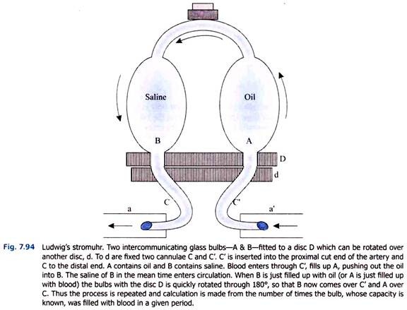
a. Ludwig’s Stromuhr (Fig. 7.94):
With this instrument the amount of blood passing through a vessel per unit time is measured. The velocity is then calculated by dividing the total volume flow (V) with the cross-sectional area of the vessel (πr2= area of circle).
b. Differential Stromuhr (Fleischl):
ADVERTISEMENTS:
This is a more accurate instrument by which the change of blood velocity during each heart beat can be recorded.
iii. Thermostromuhr (Rein):
The principle is to heat a particular spot on the vessel by high frequency current at a known rate and then the rise of temperature of blood is noted a little down the stream by a thermocouple. The rise of temperature is inversely proportional to the velocity.
iv. Electromagnetic Method:
ADVERTISEMENTS:
A more advanced method based on electromagnetic principle has also been devised.
Mean Volume Flow:
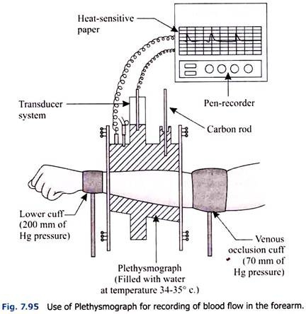
Instrument used—Plethysmograph (Fig. 7.95). The total volume of blood passing through an organ or any other part does not depend upon the blood velocity flow through the corresponding artery.
It depends upon three factors:
ADVERTISEMENTS:
1. The total cross-sectional area of the vascular bed in the organ.
2. The rate of metabolism in the organ.
3. The degree of vasodilatation or vasoconstriction in the locality.
The following figures give the mean volume flow per minute for each 100 gm:
i. Thyroid – 560 ml.
ADVERTISEMENTS:
ii. Kidney – 150 ml.
iii. Brain – 130 ml.
iv. Liver – 150 ml (1/3rd arterial, 2/3rds portal).
v. Intestine – 70 ml.
vi. Heart (Coronary) – 100 ml.
Alteration of volume of a limb is recorded by means of an instrument called Plethysrmograph (Fig. 7.95). The cuff of the Sphygmomanometer is applied to the arm and inflated up to a certain pressure which is less than the arterial pressure. The veins are occluded but the arterial blood flow in the limb remains unchanged. The volume of the limb gradually increases as no blood can leave due to occlusion of the veins.
ADVERTISEMENTS:
The limb is kept in a water-tight glass vessel which is connected with the volume recorder. Changes in the volume of the limb will displace water from the glass vessel and this displacement of water will be recorded in a sensitive electronic apparatus through transducer system.


