ADVERTISEMENTS:
The following points highlight the three main processes involved in the genetic recombination of bacteria. The processes are: 1. Conjugation 2. Transformation 3. Transduction.
Process # 1. Conjugation:
In this process, the exchange of genetic material takes place through a conjugation tube between the two cells of bacteria. The process was first postulated by Joshua Lederberg and Edward Tatum (1946) in Escherichia coli. They were awarded the Nobel Prize in 1958 for their work on bacterial genetics. Later on, it has also been demonstrated in Salmonella, Vibrio and Pseudomonas.
ADVERTISEMENTS:
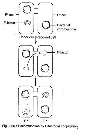
There are two mating types of bacteria, one is. male type or F+ or donor cell, which donates some DNA. The other one is female type or F– or recipient cell, which receives DNA.
Later, after receiving DNA, the recipient cell may behave as donor cell i.e., F+ type. The F-factor is the fertility factor, sex-factor or F-plasmid present in the cell of F+ i.e., donor cell or male type. The plasmid takes part in conjugation is called episome.
In this process, two cells of opposite mating type i.e., F+ and F– become temporarily attached with each other by sex pilus (Fig. 2.26). The sex pilus has a hole of 2.5 pm diameter through which DNA can pass from donor to recipient cell. The F-factor or F-plasmid is a double stranded DNA loop, present in the cytoplasm; apart from the nucleoid. The F-factor contains about 20 genes.
After the establishment of conjugation tube, the F-factor prepares for replication by the rolling circular mechanism. The two strands of F- factor begin to separate from each other and one of them passes to the recipient i.e., F– cell.
ADVERTISEMENTS:
After reaching in F– cell, enzymes synthesise a complementary strand that forms a double helix, which bends into a loop. The conversion process is thus completed. In the donor cell i.e., in F+, a new DNA strand also forms to complement the left over DNA strand of the F-factor.
There is another type of conjugation where passage of nucleoid DNA takes place through conjugation tube. Strains of bacteria are known as Hfr (high frequency of recombination) strain. William Hayes discovered such strains of E. coli in 1950s. The Hfr factor is also called episome. In Hfr strain, the F-factor is attached with the nucleoid DNA i.e., the bacterial chromosome.
In this process, Hfr and F– cells become attached with each other by sex pilus (Fig. 2.27). At the point of attachment of F-factor, the bacterial chromosome opens and a copy of one strand is formed by the rolling circular mechanism.
A portion of single stranded DNA then passes into the recipient cell through pilus. Due to agitation in medium, the conjugation tube may not survive for long time because of broken pilus. Thereby, the total length of transfer DNA may not be able to take entry to the recipient cell.
The behaviour of the transferred DNA depends on the presence and absence of F-factor:
If F-factor is indeed transferred, then it usually remains detached from the chromosome of recipient cell and enzymes synthesise a complementary DNA strand. The factor then forms a loop and exists as a plasmid, thereby the recipient cell becomes a donor.
If F-factor remains at the rear end of the transfer DNA during its entry to the recipient cell, the F-factor may not be able to take entry due to broken pilus and only a portion with new genes (Fig. 2.27) takes up the entry. Thereby, the F– strain remains as recipient one. In F– strain, genetic recombination takes place between donor fragment and recipient DNA.
ADVERTISEMENTS:
(iii) Sometimes, if the F-factor gets free from the Hfr cell and maintains an independent status, then the Hfr cell converts to a F+ cell. Sometimes during the leaving of F-factor from the bacterial chromosome, it takes a segment of chromosomal DNA. The F-factor with segment of chromosomal DNA is called F’-factor.
Later on, during conjugation, when this F’-factor is transferred, the recipient cell receives some chromosomal DNA from the donor cell. This process is called sexduction. In this process, the recipient cell receives a portion of chromosomal DNA which duplicates with the existing one for a specific function, thereby the recipient cell is a partial diploid.
Process # 2. Transformation:
It is a kind of genetic recombination where only the carrier of genes, i.e., the DNA molecules of donor cell, pass into the recipient cell through the liquid medium:
It was described by Frederick Griffith (1928), an English bacteriologist. He had done his experiment with laboratory mice and two types of Diplococcus pneumoniae, the pneumonia causing organism. One type has rough (R) non-capsulated cells and another one with smooth (S) capsulated cells. The R-type is non-pathogenic, while the S-type is pathogenic.
ADVERTISEMENTS:
The process of transformation is mentioned below (Fig. 2.28):
(i) When live non-pathogenic (R-type) cells are injected in mice, the mice remain alive.
(ii) When dead pathogenic (S-type) cells are injected in mice, the mice also remain alive.’
(iii) When pathogenic (S-type) cells are injected in mice, they suffer from pneumonia and died.
ADVERTISEMENTS:
(iv) When live non-pathogenic (R-type) cells are mixed with dead pathogenic (S-type) cells and are injected in mice, they also suffered from pneumonia and died. On isolation of dead tissue of mice, the smooth (S) qapsulated cells are found on agar. The above experiment indicates the conversion of R-type to S-type, called transformation.
Later, James L. Alloway (1932), transformed the rough type cells to smooth type, by using the fragments from dead smooth-type cells and confirmed Griffith’s work.
Further, Oswald T. Avery, Colin M. MacLeod and Maclyn N. McCarty (1944) also found that DNA isolated from the fragments could induce the transformation. Their experimental result was the first proof of DNA as the genetic material in living organism. The possible mechanism of transformation can be explained (Fig. 2.29).
The transformation takes place in a few cell of the mixed population. It is an important method of genetic recombination. A few donor cells break apart and an explosive release and fragmentation of DNA take place. A fragment of double stranded DNA (10-20 genes) then gets attached with the recipient cell for entry (Fig. 2.29).
ADVERTISEMENTS:
During entry one strand of the fragment becomes dissolved by enzyme leaving the second strand, which then passes to the recipient cell through cell wall and cell membrane.
After entry, a portion of single strand of double stranded DNA of recipient cell gets displaced by enzyme and then replaced by the DNA of donor cell. The displaced DNA is then dissolved by other enzyme. Thus the recipient cell becomes transformed which will display its own as well as the characters of the newly incorporated DNA.
Detailed mechanism of transformation, with especial emphasis on natural and induced competence and DNA uptake:
Thus the transformation takes place by horizontal gene transfer through uptake of free DNA by other bacteria. This transformation takes place either spontaneously by taking DNA from the environment, i.e., Natural, or by forced uptake under laboratory condition i.e., Artificial process.
A. Natural Transformation:
ADVERTISEMENTS:
During natural transformation, free naked fragments of double stranded DNA of donor cell become attached to the surface of the recipient cell. The free double stranded ON A molecules may be available in the medium by lysis or natural decay of bacteria (Fig. 2.30).
After attachment of donor double stranded DNA with the surface of recipient bacterium, one strand is digested by the bacterial nuclease and the remaining one strand is then taken in by an energy-requiring transport system. This uptake of DNA takes place during late logarithmic phase of growth.
During this process, Rec A type of protein plays an important role. The Rec A protein binds with the single stranded DNA and forms a coating around the DNA (Fig. 2.30). The coated single stranded DNA and DNA of recipient cell then move close to each other to get homologous sequence.
After reaching at proper place, the Rec A protein actively displaces one strand of chromosomal DNA of recipient cell. The process requires hydrolysis of ATP to get energy. The incoming DNA strand is then integrated with one strand of bacterial DNA by base pairing and ligation takes place by DNA ligase.
The displaced DNA strand of recipient cell is then digested by cellular DNase activity. Any mismatch between the two strands of new region is corrected by them. Thus the transformation is completed. If the introduced single stranded DNA fails to recombine with the recipient DNA, it is digested by cellular DNase and gets lost.
B. Artificial Transformation:
ADVERTISEMENTS:
The E. coli, an ideal material for research is not transformed naturally. Later, it has been discovered that the transformation in E. coli can be done by special physical and chemical treatments. This can be done by exposure of E. coli to high voltage electric field and also by high concentration of CaCI2. Under such condition, the bacterial cells are forced to take up foreign DNA. This type of transformation is called artificial.
During this process, the recipient bacterial cells are able to take up double stranded DNA fragments.
Physical or chemical treatment forces the recipient bacterial cell to receive exogenous DNA. The foreign DNA is then integrated with the chromosome by homologous recombination, mediated by Rec A protein. The Rec A protein catalyses the annealing of two DNA segments and exchange of homologous region.
This involves nick i.e., small cut of DNA strands and rejoining of exchanged parts i.e., breakage and reunion. The generally accepted model of the above phenomenon is given below (Fig. 2.31):
Process # 3. Transduction:
It is a special method of genetic recombination where genetic material is transferred from the donor to the recipient cell through a non- replicating bacteriophage — temperate bacteriophage. This was discovered by Joshua Leaderberg and Nortor Zinder (1952) during their research with Salrv onella typhimurium.
In this process, a small fragment of bacterial DNA is incorporated into an attacking bacteriophage (i.e., virus which infect bacteria) and when this bacteriophage infects a new bacterial cell, it transfers the genetic material into it, and thus genetic recombination takes place.
Transduction are of two types:
A. Specialised transduction, and
B. Generalized transduction.
A. Specialised Transduction:
In this process, the bacteriophage gets attached to a bacterial cell wall at the receptor site and the nucleic acid of bacteriophage is transferred into the cytoplasm of the host cell (Fig. 2.32A). The phage does not cause the lysis of the host bacterium. In the bacterial cell, the phage nucleic acid codes for the synthesis of specific proteins, the repressor proteins.
The repressor proteins prevent the virus to produce the material require for its replication. In the bacterial cell, the viral DNA may exist as a fragment in the cytoplasm or it may attach itself to the chromosome, known as prophage (Fig. 2.32B). The bacterial cell which carries the prophage is called lysogenic and the phenomenon where the phage DNA and bacterium exist together is called lysogeny.
The bacterial cell may remain lysogenic for many generations and during this period the viral DNA replicates many times together with the bacterial chromosome.
However, in course of time, the phage stops the synthesis of repressor proteins in the bacterial cell, and then the synthesis of phage components starts. Now the phage DNA separates from the bacterial chromosome and starts the synthesis of phage proteins (Fig. 2.32C).
During this separation, a number of genes of the bacterium get attached to it. These attached genes keep on replicating along with the phage DNA (Fig. 2.32D) and later on it develops into phage particles, those come out from the bacterial cell by bursting (Fig. 2.32E).
When the new phage particle (Fig. 2.32F) infects a new bacterial cell (Fig. 2.32G, H), the attached bacterial genes present along with phage particle enters in the chromosome of the new bacterium and causes recombination (Fig. 2.321).
Thus the new bacterial cell contains its own genes and several genes from the parent bacterial cell. This type of transduction is known as specialised transduction, which is an extremely rare event.
B. Generalised Transduction:
This process of transduction is more common than specialized transduction. Here the prophage particle is present in the cytoplasm of the infected bacterial cell (Fig. 2.32J). In this process, the phage DNA starts synthesising new phages.
During this process chromosome of bacterial cell gets fragmented (Fig. 2.32K) and some of the fragments become attached with the DNA of some new phage particle, while others remain with phase DNA (Fig. 2.32L).
When the newly formed phage with fragment of bacterial chromosome in its DNA (Fig. 2.32M) attacks a new bacterium, the gene of the parent bacterium is transferred to the new bacterium and causes recombination. This type of transduction is called generalised transduction. This type of transduction is also rare.


