ADVERTISEMENTS:
In this article we will discuss about the anatomy of different normal monocot stems: 1. Zea mays- Stem 2. Canna – Stem 3. Triticum – Stem.
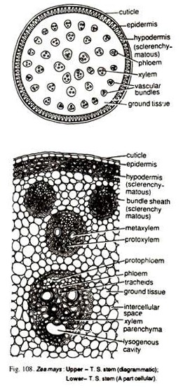
1. Anatomy of Zea mays – Stem (Family – Graminae):
T.S. exhibits following tissues from outside within:
It is circular in outline with a well-defined epidermis, hypodermis, ground tissue and many scattered vascular bundles.
ADVERTISEMENTS:
Epidermis:
1. It is the outermost layer of stem.
2. The outer wall of cells is covered by a thick cuticle.
3. The continuity of the layer is broken by few stomata.
ADVERTISEMENTS:
4. Epidermal hairs are absent
Hypodermis:
5. It is two to three cells thick, sclerenchymatous and present just below the epidermis.
6. Cells are polygonal in shape.
Ground tissue:
7. It is not differentiated into cortex, endodermis, pericycle and pith.
8. The cells are parenchymatous and extend from below the sclerenchyma to the centre.
9. The cells are small and compactly arranged below the hypodermis but they are large, round and loosely arranged in the centre.
Vascular Bundles:
ADVERTISEMENTS:
10. Vascular bundles are many and scattered in the ground tissue with no definite arrangement.
11. They are small and more in number towards the periphery than the centre of the section.
12. Each vascular bundle is conjoint, collateral, closed and endarch.
13. A well developed sclerenchymatous sheath surrounds each vascular bundle which is more prominent at its upper and lower faces.
ADVERTISEMENTS:
14. Xylem and phloem constitute the vascular bundle.
15. Phloem:
(i) Consists of only sieve tubes and companion cells.
(ii) Phloem fibres and phloem parenchyma arc absent.
ADVERTISEMENTS:
(iii) The outer parts of the phloem, which is broken and disorganized, is called protophloem.
(iv) Inner phloem contains sieve tubes and companion cells, and called metaphloem.
16. Xylem:
(i) Consists of vessels (protoxylem and metaxylem), tracheids and xylem parenchyma.
ADVERTISEMENTS:
(ii) Vessels are in the from of ‘Y’.
(iii) Metaxylem is present at the divergent ends of ‘Y’ in the form of two big oval vessels.
(iv) Protoxylem is present at the lower arm of ‘Y’, consisting of two small vessels.
(v) Protoxylem is surrounded by tracheids and xylem parenchyma.
(vi) Inner protoxylem vessel and parenchyma break down and form a water-containing cavity called lysigenous cavity.
Identification:
ADVERTISEMENTS:
(a) 1. Presence of vessels in the xylem. (Angiosperms)
(b) 1. Vascular bundles are conjoint, collateral and endarch. (Stem)
(c) 1. No differentiation of ground tissue.
2. Sclerenchymatous hypodermis.
3. Vascular bundles are closed.
4. Scattered vascular bundles.
ADVERTISEMENTS:
5. Absence of secondary growth. (Monocot)
Special Points:
1. Scattered vascular bundles.
2. ‘Y’ -shaped vessels.
3. Presence of protophloem and metaphloem.
2. Anatomy of Canna – Stem (Family – Cannaceae):
T.S. reveals the following tissues from outside within:
It is circular in outline with a well-defined epidermis, hypodermis, ground tissue system and many scattered vascular bundles.
Epidermis:
1. It consists of many small, flat and tangentially elongated cells.
2. A thick cuticle covers the outer wall of the cells of epidermis.
3. Epidermal hairs are absent.
Ground Tissue System:
4. It consists of cortex, chlorenchyma, patches of sclerenchyma and ground tissue.
5. Just below epidermal layer are present two layers of cortex, consisting of large polygonal cells.
6. Chlorenchyma is present immediately below the cortex in the form of one or two layers.
7. Sclerenchyma patches remain attached with the chlorenchyma.
8. Rest of the portion is filled with many large, thin walled, parenchymatous cells which form ground tissue.
9. Large intercellular spaces are present in the ground tissue.
Vascular Bundles:
10. Many vascular bundles are scattered in the ground tissue.
11. Vascular bundles are of different sizes.
12. Each vascular bundle is conjoint, collateral, closed and endarch.
13. Each vascular bundle is covered by incomplete, sclerenchymatous bundle sheath. It is rarely complete.
14. Bundle sheath is present in the form of a large patch on the outer side and a small strip on the inner side of vascular bundle.
15. Each vascular bundle is made up of phloem and xylem.
16. Phloem is situated towards the outer side in the vascular bundle and consists of companion cells and sieve tubes.
17. Xylem is situated towards the inner side in the bundle, and consists of few large and small vessels and xylem parenchyma.
Identification:
(a) 1. Presence of vessels in the xylem. (Angiosperms)
(b) 1. Vascular bundles are conjoint, collateral and endarch. (Stem)
(c) 1. Well-developed ground tissue.
2. Scattered vascular bundles.
3. Vascular bundles are closed.
4. Absence of secondary growth. (Monocot)
Special Points:
1. Incomplete bundle sheath.
2. Presence of sclerenchymatous patches in the ground tissue.
3. Anatomy of Triticum – Stem (Family – Graminae):
Following tissues are visible in the T.S. of the material from outside within.
It is circular in outline with a layer of epidermis, ground tissue, vascular bundles and a well developed hollow cavity in the centre.
Epidermis:
1. The epidermis is single layered and consists of rectangular cells.
2. A thick cuticle is also present
3. Continuity of epidermis is broken by some stomata.
Ground Tissue-System:
4. It is made up of sclerenchyma, chlorenchyma and parenchymatous ground tissue, and occupies the major portion of the stem.
5. Below the epidermis is present a zone of sclerenchyma which is many cells deep.
6. The regularity of the sclerenchyma is interrupted by patches of chlorenchyma.
7. The stomata open into a sub-stomatal cavity only in the region of chlorenchyma.
8. The remaining part of the ground tissue system consists of thin walled parenchymatous cells.
9. Many intercellular spaces are present in the parenchymatous region.
Vascular Bundles:
10. Many vascular bundles are present in the ground tissue.
11. Vascular bundles are arranged in two series.
12. The bundles of the inner series are larger than that of outer series.
13. In majority of the Cases the bundles of the outer series remain embedded in the sclerenchyma of the ground tissue.
14. Vascular bundles are conjoint, collateral, closed and endarch.
15. Each vascular bundle remains surrounded by a definite bundle sheath.
16. Each vascular bundle consists of xylem and phloem.
17. The xylem is located in the lower region of the vascular bundle, and is Y-shaped in its organization.
18. The metaxylem elements are large and present towards the outer side whereas the protoxylem elements are smaller and endarch.
19. The phloem consists of sieve tubes and companion cells.
Pith Cavity:
20. The centre is occupied by a large, well developed, hollow pith cavity.
Identification:
(a) 1. Presence of vessels in the xylem. (Angiosperms)
(b) 1. Vascular bundles are conjoint, collateral and endarch. (Stem)
(c) 1. Well developed ground tissue.
2. Vascular bundles are closed.
3. Absence of endodermis and pericycle.
4. Bundle sheath well developed.
5. Absence of secondary growth. (Monocot)
Special Points:
1. Vascular bundles are almost in two rings.
2. A large, hollow pith cavity.
3. Majority of the bundles of the outer series are embedded in the sclerenchyma.


