ADVERTISEMENTS:
In this article we will discuss about the external and internal anatomy of earthworm. This will also help you to draw the structure and diagram of earthworm.
External Anatomy of Earthworm:
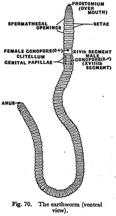
The body of Pheretima is nearly circular in cross-section and varies from 7 to 8 inches (18-19 cms) in length. The general colour of the body is brown but the dorsal surface is darker. A dark line extends from end to end in the mid-dorsal line.
The anterior end of the body is pointed and the posterior end is blunt. The animal is elongated and divided into a series of ring-like segments or metameres or somites, which are separated from one another by narrow transverse grooves.
ADVERTISEMENTS:
Usually there are 120 true somites. A fleshy lobe, the prostcmium, projects over the mouth in front of the first segment. The skin of segments, fourteenth, fifteenth and sixteenth, is swollen and pale in mature worms to form a saddle-shaped structure called clitellum or cingulum; it secretes the material for producing cocoons.
Every segment of the body, excepting the clitellar, and the first and last, bears numerous chitinous bristles called setae; these are implanted on the body wall and are arranged in the form of rings. The setae serve as hold-fasts when the worm is moving over the ground or resting in its burrow.
The entire body is covered by a thin transparent cuticle which is secreted by the epidermis. The cuticle is porous and protects the body from injury.
A number of external openings are found in the body to carry on different functions:
ADVERTISEMENTS:
(1) The mouth is a small crescentic aperture situated at the anterior end of the first segment just beneath the prostomium. It is meant for ingestion of food.
(2) The oval anal aperture for the exit of faeces lies at the posterior end in the last segment.
(3) The paired male gonopores are situated ventrally on the eighteenth segment, one on each side of the mid-ventral line. These are the openings of the sperm-ducts.
(4) The genital papillae are two pairs of cup-like depressions on the ventral surface of the seventeenth and nineteenth segments. The two papillae of a side, are placed one in front of and the other behind the corresponding male gonopore.
(5) The single female gonopore is a median aperture on the ventral side of the fourteenth segment; it serves as an exit for the eggs.
(6) Four pairs of spermathecal apertures are situated in the inter-segmental grooves between Vth and VIth, Vlth and VIIth, Vllth and VIIIth, and Vlllth and IXth somites. Each of these leads into a pouch called spermatheca which acts as the receptacle of sperms received from another worm at the time of mating.
(7) The body cavity or coelom communicates with the exterior through dorsal pores. These occur in the inter-segmental grooves along the mid-dorsal line. The first dorsal pore is in the groove between the XIIth and XIIIth segment, and there is one in each subsequent inter-segmental groove, excepting the last.
(8) The nephridiopores or the apertures of the excretory organs are minute openings found scattered all over the body, accepting the first three segments and the last.
Internal Anatomy of Earthworm:
If a worm is cut open from the anterior to the posterior end by an incision through the body wall in the mid-dorsal line, the internal structures may easily be studied. It is to be noted that the body of the earthworm is essentially a double tube. The body wall is the outer tube and the alimentary canal is the inner tube. The two tubes are separated by an extensive space, the body cavity or coelom.
ADVERTISEMENTS:
External segmentation of the body corresponds to internal partition of the coelom into compartments by a number of septa which lie beneath the inter-segmental grooves. Thus, the body cavity is divided into 100 or more compartments corresponding to the number of external somites.
Each compartment is lined internally by epithelium and is filled with a milky fluid which flows from compartment to compartment, because each septum is perforated by a large opening.
The alimentary canal remains suspended in the coelom and is held in position by the septa, which appear to arise from its wall. As the first septum lies between the IVth and Vth segments the coelom anterior to this region is a continuous cavity.
ADVERTISEMENTS:
The milky coelomic fluid may spurt out through the dorsal pores when the worm is irritated. It contains large granular phagocytes, small yellow cells, and a few circular nucleated cells of intermediate size. It keeps the skin of the worm moist, and helps in the excretion of waste products. The phagocytes devour bacteria which are injurious to the earthworm, and thus protect it.
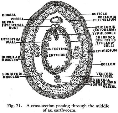
Running along the dorsal surface of the alimentary canal is the dorsal blood vessels, beneath which is the supra-intestinal duct. There is a ventral blood vessel ventral to the gut and overlying the ventral nerve cord. Beneath the ventral nerve cord is a small sub-neural vessel.
The reproductive organs are suspended in the coelom and extend from the Vlth to the XlXth segments. The body wall and the gut wall are best seen in a cross-section passing through the middle of the worm (Fig. 71).
The body wall is composed of the following layers:
ADVERTISEMENTS:
(i) The outermost layer is the thin and transparent cuticle which is a porous non-cellular membrane covering the epidermis. The epidermis secretes and replaces the cuticle from time to time.
(ii) The epidermis is composed of a single row of epithelial cells. Some of these are modified into gland cells secreting mucus which is poured out through the cuticle and cleanses the body surface. A few epidermal cells are modified as receptors and act as sense organs. Others are merely supporting cells. These are sacs in the epidermis for implantation of the setae, and minute muscle fibres for moving them.
(iii) Beneath the epidermis, there is the musculature of the body which consists of an outer circular layer and an inner longitudinal layer. The former produces a continuous sheath running round the body and the latter runs in parallel bundles along the length of the worm. Pigment cells and blood capillaries are scattered among the muscle fibres.
ADVERTISEMENTS:
(iv) A thin layer of coelomic epithelium, consisting of a single row of flat cells, lies just beneath the longitudinal muscles. It forms the innermost lining of the body wall. The body wall is not only protective but it also carries on respiration through its moist outer surface. The musculature along with the setae are responsible for locomotion.
The contraction of the circular muscles elongates, whereas contraction of the longitudinal muscles shortens the length of the body. The two kinds of muscles are brought into play alternately. The setae are used for fixation on the substratum. The skin contains the receptor organs and therefore serves as a sensory membrane.
The got wall is composed of the following layers:
(I) An outer serous coat formed by a layer of tall narrow cells. They are seen to be loaded with minute yellowish granules, specially in the intestinal region of the gut, and are therefore called chloragogen cells or yellow cells. They collect waste products, and when fully loaded, drop down into the coelom and are discharged through the dorsal pores, thus helping in excretion.
(II) Beneath the serous coat are two layers of muscles—the outer one being longitudinal and the inner one circular in disposition. The musculature is poorly developed and consists of non-striped fibres. In the region of the oesophagus and the gizzard the musculature is well-formed.
ADVERTISEMENTS:
(Ill) The internal lining of the gut is a single layer of epithelial cells some of which are glandular and others are absorptive. The glandular cells secrete digestive juice.


