ADVERTISEMENTS:
In this article we will discuss about:- 1. Occurrence of Oedogonium 2. Plant Body of Oedogonium 3. Features 4. Cell Structure 5. Reproduction 6. Life Cycle.
Occurrence of Oedogonium:
Oedogonium (Gr. oedos, swelling; gonos, reproductive bodies) is an exclusively fresh water alga. Out of about 400 species more than 200 have been reported from India. They are very common in pools, ponds, lakes etc.
The filamentous plant body may get attached with the stone, wood, leaves of aquatic plants, small branches of dead plant remain in water etc. by their basal cell the holdfast. Some species like O. terrestris are terrestrial.
Plant Body of Oedogonium:
ADVERTISEMENTS:
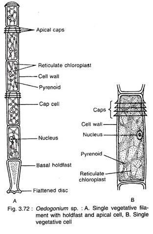
The thalloid plant body is green, multicellular and filamentous. The filaments are unbranched and cells of each filament are attached end to end and form uniseriate row (Fig. 3.72A). The filament is differentiated into 3 types of cells: 1. Basal cell, 2. Apical cell and 3. Middle cells.
1. Basal Cell:
It is the lowermost cell of the filament. The cell is long, gradually narrowed and towards the basal end it expands to form simple, disc-like, multilobed or finger-shaped structure. The cell is generally colourless, which performs the function of fixation to the substratum and called holdfast.
ADVERTISEMENTS:
2. Apical Cell:
It is the topmost cell of the filament. The cell is usually rounded towards apical side and green in colour.
3. Middle Cells:
All the cells in between basal and apical cells are alike. The cells are longer than their breadth i.e., rectangular in shape.
Towards the upper end of some cells a ring-like structure is present known as cap or apical cap (Fig. 3.72A). The cell with cap is called cap cell. The number of caps on a cell indicates the number of cell divisions in that cell.
Important Features of Oedogonium:
1. This is a common fresh water alga growing on substratum like sand particles, rocks etc.
2. The plant body is unbranched, filamentous and differentiated into apex and base.
3. Cells have reticulate chloroplasts.
4. Presence of caps on the young dividing cells.
ADVERTISEMENTS:
5. Vegetative cell division is very elaborate.
6. Asexual reproduction takes place by multi- flagellate zoospore, where flagella are arranged around the beak-like apical region.
7. Sexual reproduction is advanced oogamous type.
8. The female gamete i.e., ovum, is produced singly in each oogonium.
ADVERTISEMENTS:
9. The male gametes i.e., antherozoids, are very much similar to zoospores but smaller in size. Two antherozoids are produced in each antheridium.
10. Based on the size of male filament the plants are divided into two groups: Macrandrous and Nannandrous type.
11. In macrandrous type the antheridia develop into the filament of normal size. But in nan- nandrous type the antheridia develop on small and thin male filament, the dwarf male or nannandrium (remain attached with the oogonium wall or its lower cell, the supporting cell), develop on germination of andro- spore. The androspore forms singly in andro- sporangium, develop in the normal filament.
12. The androspores are smaller than zoospores (produced asexually), but larger than antherozoids.
ADVERTISEMENTS:
13. The zygote undergoes meiotic division and produces four zoospores. In dioecious species two produce male and other two produce female plants.
Cell Structure of Oedogonium:
The intercalary cells are longer than their breadth and are cylindrical in outline.
The cells are surrounded by thick and rigid cell wall (Fig. 3.72B). The cell wall is differentiated into three layers an outer chitin, middle pectin and innermost cellulosic. Just interior to the wall, cell membrane is present, which encloses the protoplast.
The protoplast consists of cytoplasm, chloroplast and nucleus. The cells contain many small or single large vacuoles situated in the centre and remain filled with cell sap. The cytoplasm lies between the cell membrane and vacuole.
ADVERTISEMENTS:
The Chloroplast is single, large and reticulate, which remains embedded in the cytoplasm. It extends from one end of the cell to the other end. Cells are uninucleate and nucleus is generally present in the centre of the cell within the cytoplasm or it may be excentric.
Cell Division:
Growth of the filament takes place through cell division (Fig. 3.73). All cells except apical and basal ones are capable of dividing through cell division though division remains restricted in some of the cells of the filament.
The steps of cell division are:
1. Initially the nucleus becomes shifted from peripheral position towards the centre and then moves slightly towards the upper half of the cell (Fig. 3.73A, B).
ADVERTISEMENTS:
2. Ring-like thickening develops towards the upper part of the cell wall which gradually increases in thickness (Fig. 3.73B).
3. The nucleus undergoes mitotic division and form two nuclei (Fig. 3.73C).
4. At the end of cell division (telophase), a row of microtubules develop and accumulate as a layer between the daughter nuclei (Fig. 3.73C). This layer remains in floating condition which will develop the future septum.
5. The ring-like thickening gradually elongates and splits the mother wall towards the apical region. The ring expands much more and forms a concave cylindrical structure (Fig. 3.73D). The ring material ultimately forms the cuticle of the upper daughter cell.
6. The upper part of the ruptured mother wall remains attached to the anterior end of the new daughter cell as a cap i.e., the apical cap. The other part remain towards the basal region of the daughter cell (Fig. 3.73D).
7. The floating septum gradually goes up to the base of the future daughter cell i.e., at the top of the mother cell at the ruptured end and it becomes fixed (Fig. 3.73E). Later on it develops into mature cross wall.
ADVERTISEMENTS:
8. New side wall develops between the cuticle and the plasmalemma of the upper cell. Thus the two cells are formed (Fig. 3.73F). It is evident that the cell with cap is the younger one which develops between the two old cells.
Reproduction in Oedogonium:
Oedogonium reproduces by all the three means: vegetative, asexual and sexual.
Vegetative Reproduction:
It takes place by fragmentation and akinete formation:
1. Fragmentation:
It takes place by accidental breakage of the filament, dying off of intercalary cells or by the formation of intercalary sporangia. The fragments are capable of developing into new filaments.
2. Akinete:
During unfavourable condition the entire protoplast of a cell becomes a thick-walled, reddish-brown, round or oval structure, the akinete. The akinete germinates during favourable condition and develops a new filament. They generally form in chain.
Asexual Reproduction:
Asexual reproduction takes place by means of zoospores (Fig. 3.74A-C). Zoospores are formed singly within a cell. Comparatively younger cell i.e., the cell with cap behaves as sporangium mother cell.
The zoospores are multiflagellate and ovoid, pyriform or spherical in shape. They are uninucleate with single chloroplast and occasionally with an eye-spot.
During favourable condition, the zoospore formation begins in a cap cell of the filament. The entire protoplast of zoosporangium contracts from the wall and behave as a unit. The protoplast becomes round or oval in shape and its nucleus moves at one end.
Near the nucleus a semicircular hyaline area develops. Just below the hyaline area a ring of blepharoplast granules develops, connected with each other by fibrous strands (Ringo, 1967). Later on, from each blepharoplast granule, single flagellum develops. Thus a crown of flagella is present around the colourless semicircular area.
The fully developed zoospores are liberated by breaking the zoosporangium wall. The wall of the zoosporangium breaks near the cap region and the neighbouring cell bend on one side to make way for the liberation of zoospore. During liberation, the zoospore remains as a delicate mucilaginous vesicle for 3-10 minutes. After dissolution of vesicle the zoospore gets free and starts swimming in the surrounding water.
Germination:
The zoospore can swim for about one hour or more. Coming in contact with substratum by the anterior end, it loses flagella and starts to elongate. The lower hyaline part becomes separated by cell wall, which forms the hold fast. Through the subsequent division and re-division in a single plane, new filament is formed.
Sexual Reproduction:
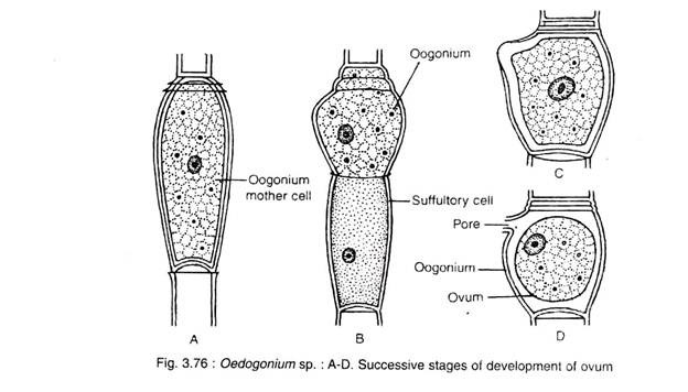
The sexual reproduction in Oedogonium is an advanced oogamous type. The male gametes or antherozoides are produced in antheridium (Fig. 3.75) and the female gamete or egg is produced in oogonium (Fig. 3.76). Male and female gametes differ both morphologically and physiologically.
Only one egg is produced in each oogonium and two antherozoides in each antheridium. Another motile structure, the androspore, is produced singly in each androsporangium. Deficiency of nitrogen and alkaline pH are the important factors for promoting sexual reproduction.
Distribution of Sex Organ in Oedogonium:
Based on the size of the male (antheridial) filament the species of Oedogonium are divided into two groups macrandrous and nannandrous type:
1. Macrandrous Type:
In macrandrous type the antheridium develops in the filament of normal size.
It is of two Types:
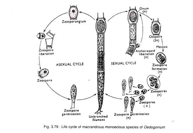
i. Monoecious type (homothallic or bisexual). In this type (e.g., O. fragile, O. nodulosum and O. hirnii) antheridia and oogonia are borne on the same filament (Fig. 3.79).
ii. Dioecious type (heterothallic or unisexual). In this type (e.g., O. gracilius, O. cardiacum and O. aquaticum) the antheridia and oogonia are borne on the different filaments (Fig. 3.80).
2. Nannandrous Type:
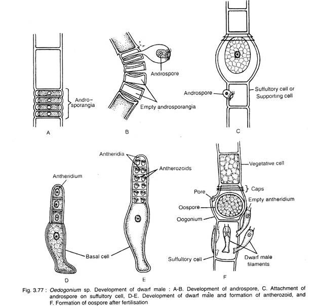
The nannandrous species are always dioecious (heterothallic) i.e., antheridia and oogonia are borne on different filaments. In this type the antheridia develop on a very small filament termed as dwarf male or nannandrium. In nannandrous type initially androsporangia are developed in series on normal sized filament. The androspore form singly within androsporangium.
Liberating from androsporangium, the androspores swim freely in water. The androspore germinates on the oogonial wall (O. ciliatum) or on supporting cell (O. concatenatum) and forms dwarf male filament. Towards the apical region, the dwarf male filament cuts off small cells as the antheridial mother cells.
Each antheridium produces two antherozoides. The androspores, antherozoids and zoospores are morphologically alike but differ in their sizes (Table 3.1). The androspores are smaller than zoospores (produced asexually) but larger than antherozoides.
They are of two types:
i. Cynandrosporous Type:
In this type (e.g., O. concatenatum) the androsporangia and oogonia are borne on the same filament (Fig. 3.81).
ii. Idioandrosporous Type:
In this type (e.g., O. setigerum, O. confertum and O. iyengarii) the androsporangia and oogonia are borne on different filaments (Fig. 3.82).
Sexual Reproduction in Macrandrous Species:
The structure and development of antheridium and oogonium are similar in all the species belonging to either monoecious or dioecious type. They differ only in the position of sex organs. In monoecious type both the sex organs develop on the same filament, but in dioecious type they are on different filaments.
a. Antheridium:
Any cap cell of the vegetative filament may function as antheridial mother cell (Fig. 3.75). It divides transversely into an upper smaller antheridium and a lower larger sister cell. The sister cell then undergoes repeated transverse division and form an uniseriate row of about 2-40 rectangular uninucleate antheridia.
The nucleus of the antheridium undergoes mitotic division and forms 2 nuclei. Each nucleus becomes surrounded by some cytoplasm and metamorphoses into an antherozoid. Thus two antherozoids are developed from each antheridium.
The antherozoids are unicellular, uninucleate, multiflagellate and yellowish in colour. Morphologically it is similar to zoospore and androspore, but much smaller in size. The liberation of antherozoid is similar to zoospore formed during asexual process.
b. Oogonium:
Any cap cell of the vegetative filament may function as oogonial mother cell (Fig. 3.76). It divides transversely into an upper oogonium and a lower supporting cell or suffultory. The lower cell may again undergoes similar divisions in repeated sequence to form two or more oogonia with a lower supporting cell.
With maturity the oogonium becomes globose, which contains single egg. A receptive spot is present at one side of the egg. Before fertilisation a transverse slit or pore develops on the oogonial wall through which the antherozoids take the entry.
Sexual Reproduction in Nannandrous Species:
The structure and development of androsporangium, antheridium and oogonium are similar in all the species either belonging to Gynandrosporous or Idioandrosporous type. They differ only in the position of androsporangium. In Gynandrosporous type the androsporangia and oogonia are borne on the same filament, whereas in Idioandrosporous type the androsporangia and oogonia are borne on different filaments.
a. Androsporangium:
The mode of development of androsporangia is alike with the antheridial development in macrandrous species. The androsporangia are larger than the antheridia of macrandrous type. The nucleus of androsporangium does not divide and the entire protoplast metamorphoses into a single androspore.
The androspores are unicellular, uninucleate and multiflagellate. The androspores are larger than the antherozoids. The androspores are liberated by breaking the wall of androsporangium. During liberation each androspore remains in a mucilage envelop for few minutes and then becomes free to swim in water (Fig. 3.77A, B).
b. Germination of Androspore and Formation of Antherozoids:
After swimming for some time, it gets attached either on oogonial wall or on supporting cell (Fig. 3.77C). Then a wall develops around the andospore. The androspore elongates and cuts off a few flat cells at its apex to form the antheridia (Fig. 3.77D). The nucleus of each antheridium divides mitotically to form two nuclei.
Each nucleus with some cytoplasm metamorphoses into single antherozoid. Thus two antherozoids are formed in each antheridium (Fig. 3.77E). The antherozoids are liberated in a similar way as found in macrandrous species. The antherozoids swim in water for sometime and in contact with receptive pore or slit, antherozoid enters inside the oogonium and fertilizes the egg.
c. Oogonium:
The structure and development of oogonium are same as macrandrous species.
Fertilisation:
Antherozoids are attracted by the mature oogonium through chemical stimulus. Normally only one antherozoid enters through the opening on the oogonial wall and fertilises the egg, resulting in the formation of a diploid zygote or oospore (Fig. 3.78A, B; 3.77F).
Oospore:
The zygote during further development retracts itself from the oogonial wall and secretes 2-3 layered outer wall (Fig. 3.78B). Later on the outermost one becomes ornamented. The zygote generally undergoes a long period of rest and becomes brown in colour.
Germination of Oospore:
The oospore germinates during favourable condition (Fig. 3.78C-G). The nucleus undergoes meiosis and forms 4 haploid daughter nuclei. The nuclei accumulate some cytoplasm and form 4 daughter protoplasts. They liberate by rupturing the oospore wall. During liberation they develop flagella and are called meiospores or zoomeiospores.
Initially they remain inside a delicate vesicle, which soon disintegrates and the zoospores get free into the environment. After swimming for some time in water they withdraw their flagella and germinate into new haoloid Oedogonium filament like zoospore in asexual reproduction. The nature of zoomeiospore development varies in monoecious and dioecious species.
In monoecious species all the zoomeiospores develop into similar Oedogonium filament.
In dioecious species out of 4 zoomeiospores, 2 develop into male and other 2 develop into female Oedogonium filaments.
Indian Species:
Oedogonium cardiacum, O. aster, O. elegans, O. aerolatum and O. armigerum.
Life Cycle of Oedogonium:
Fig. 3.79-3.82 depict life cycle of Oedogonium.


