ADVERTISEMENTS:
In this article we will discuss about the vegetative, asexual and sexual methods of reproduction that occur in the life cycle of oedogonium.
(i) Vegetative Reproduction:
Vegetative reproduction takes place by fragmentation and akinete formation.
(A) Fragmentation:
ADVERTISEMENTS:
Oedogonium filament breaks into many small fragments which have capability to grow into complete filaments under favourable conditions.
Fragmentation takes place due to any of the following reasons:
(a) Accidental breaking of the filaments.
(b) Dying or dehydration of intercalary cells.
ADVERTISEMENTS:
(c) Disintegration of intercalary cells due to conversion in sporangia.
(d) Mechanical injury to the filament.
(e) Change in the environmental conditions.
(B) Akinete formation:
The akinetes are formed under unfavorable conditions. Akinetes are modified vegetative cells which become swollen, round or oval, reddish brown and thick walled. These are rich in reserve starch and orange-red coloured oil. Akinetes are formed in chains of 10 to 40 (Fig. 4). Akinetes germinate directly under favourable conditions.
(ii) Asexual Reproduction:
ADVERTISEMENTS:
Asexual reproduction takes place by means of multi-flagellate zoospores produced singly in intercalary cap cell. Mostly the newly formed cap cell functions as the zoosporangium.
Several factors control zoospore formation of which high pH and CO2 concentration of medium and a diurnal rhythm of light and darkness are significant. The zoospores are not formed in chains and one sterile cell is always present between two zoosporangia.
The cell which functions as zoosporangium gets filled with abundant reserve food and a slight contraction of the protoplast from the cell wall takes place (Fig. 5 A, B).
The central vacuole disappears the chloroplast frees itself from one end of the cell and becomes conical. The nucleus comes to lie near this chloroplast. A small lens shaped hyaline region is formed between the wall and the nucleus. This hyaline bald spot later forms the anterior end of the zoospore.
ADVERTISEMENTS:
At the base of this hyaline area a ring of basal granules appears and from each basal granule or blepharoplast a flagellum arises. The basal granules are connected to each other by fibrous strand. A crown of about 30 flagella is formed around the hyaline spot (Fig. 5 C).
The mature zoospore is oval, spherical or pear shaped structure. The zoospore is uninucleate and contains a ring shaped chloroplast. The zoospore is dark green in colour except at the hyaline pointed apical end. A sub apical ring of flagella is present and such flagellation is called stephanokontic type (Fig. 5 F).
When the zoospore is mature, the wall of the zoosporangium splits near the apical region and the adjacent cell moves apart to make a gap for the liberation of zoospore (Fig. 5 D).
The mucilage substance is secreted at the base of the zoospore which helps in the liberation of zoospore. The zoospore comes out of the zoosporangium in a delicate mucilaginous vesicle which soon gets dissolved and the zoospores are liberated in water (Fig. 5 D, E).
ADVERTISEMENTS:
Germination of Zoospore:
After liberation, the zoospore swims for about an hour. Then it settles and attaches itself to a solid substratum with its anterior end downwards. After attachment flagella are withdrawn and it starts elongation. The lower hyaline part elongates to make holdfast and the upper part divides repeatedly to make new filament (Fig. 5 G-I).
(iii) Sexual Reproduction:
The sexual reproduction in Oedogonium is of advanced oogamous type. Sexual reproduction is more frequent in still waters than in running water. The factors influencing sexual reproduction are alkaline medium, deficiency of nutrition, light and dark periods and increased temperature.
ADVERTISEMENTS:
The genus Oedogonium exhibits sexual dimorphism because the male and the female gametes differ morphologically as well as physiologically. The male gametes are produced in antheridia and the female gametes are produced in oogonia.
Depending upon the nature of antheridia producing plants, Oedogonium species are of two types:
(i) Macrandrous:
If antheridia are produced on normal size plant, Oedogonium forms are called macrandrous. Macrandrous species may be monoecious or dioecious. In monoecious macrandrous species antheridia and oogonia are produced on the same plant e.g., O. fragile, O. hirnii, O. kurzii and O. nodulosum. In dioecious macrandrous species antheridia and oogonia are produced on separate male and female plants of normal size.
(ii) Nannandrous:
The female or oogonia bearing plants are normal. The antheridia are produced on special type of small or dwarf plants, known as Dwarf males or Nannandria. The dwarf males are formed by androspores which are produced in androsporangia.
ADVERTISEMENTS:
If androsporangia and oogonia are formed on same plant, the Oedogonium forms are called gynandrosporous e.g., O. concatinatum. If androsporangia and oogonia are formed on different plants, Oedogonium forms are called idioandrosporous e.g., O. confertum, O. iyengarii and O. setigerum. According to some algologists, nannondrous species are more primitive.
Antheridia:
(i) In macrandrous forms:
The antheridia develop on normal filaments, terminal or intercalary in position. The initial cell which gives rise to antheridia is called antheridial mother cell. It is normally a cap cell. The antheridial mother cell divides by transverse division to form an upper smaller cell called antheridium and a lower larger cell called sister cell.
The sister cell divides repeatedly to form a row of 2-40 antheridia (Fig. 6 A). Rarely the antheridia are produced singly. The antheridia are broad, flat, short cylindrical, uninuleate cells. The contents of an antheridial cells divide either longitudinally or transversely into two.
The two antherozoids are positioned side-by-side or one above the other if divisions are longitudinal and transverse respectively. The antherozoids are liberated in the same fashion as zoospores (Fig. 6 B). The liberated antherozoids or spermatozoids or sperms are pale green or yellow green, oval or pear shaped.
ADVERTISEMENTS:
The antherozoids are motile about 30 sub-apical flagella present at the base of beak or hyaline spot (Fig. 6 C). The flagella are sometimes longer than the body of spermatozoid e.g., in O. crassum and O. kurzii. The antherozoids swim freely in water before they reach oogonia and take part in fertilization. The antherozoids are similar to zoospores in structure but these are smaller than zoospores.
(ii) In nannandrous forms:
The antheridia are formed on short or dwarf male plants called dwarf males or nannandria (Fig. 7 G). The dwarf male filament is produced by the germination of a special type of spore known as androspore.
The androspore is produced singly within an androsporangium. Androporangia are more or less similar looking to the antheridia of macrandrous forms and are produced in a similar manner from a mother cell (Fig. 7 A, B).
The androsporangia are flat, discoid cells slightly larger than antheridia. Each androsporangium produces a single androspore just as in the case of zoospore. Liberation of androspore is similar to that of a zoospore. The androspores look similar to zoospore except for the smaller size. The androspores are motile and have a subpolar ring of flagella.
After swimming about for some time, the androspore settles on oogonial wall e.g., O. ciliatum or on the supporting cell e.g., O. concatenatum. The androspore germinates into a dwarf male or nannandrium. Germlings at one celled stage may divide and produce two antherozoids e.g., O. deplandrum, O. perspicuum (Fig. 7 C-G).
The nannandrium or dwarf male can be a few cells long. It has a basal attaching cell the stipe and all others cells are antheridial cells. In many cases cap is present at the top of the apical antheridium. The protoplasm of each antheridial cell divides to form two sperms or antherozoids which are similar to antherozoids of macrandrous species.
According to Iyengar (1951) the antheridium of nannandrium produces single antherozoid. The antherozoids are released by disorganization of antheridial cell or through the opening. Pascher considered the nanandrous forms as primitive and macrandrous as specialized but a large number of phycologists consider that nannandrous species have been evolved from macrandrous species.
Oogonia:
In Oedogonium the female sex organ oogonia are highly differentiated female gametangia. These are mostly intercalary but sometimes can be terminal e.g., O. palaiense.
The structure and development of oogonium is identical in macrandrous and nannandrous species. Like antheridia any freely divided or actively growing cap cell functions as the oogonial mother cell. The oogonial mother cell divides by transverse division into two unequal cells, the upper cell and the lower cell.
The upper larger cell forms oogonium and the lower smaller cell function as supporting cell or suffultory cell. In some species the oogonial mother cells directly forms the oogonium. Supporting cell is absent is O. americanum. If any of the two divided cells again functions as oogonial mother cell many oogonia are formed in chain.
In monoecious species the suffultory cell may divide to form antheridia. The upper cell contains more cytoplasm, food and enlarges into spherical or flask shaped oogonium. The oogonium also secretes growth hormones which induce suffultory cell to increase in size (Fig. 8 A-C).
The protoplast in oogonium metamorphosis’s into a single egg or oosphere. The oosphere is non-motile, green due to chlorophyll and has a central nucleus. As the ovum matures, the nucleus moves to periphery, the oosphere retracts slightly from the oogonial wall and develops a hyaline or receptive spot just outside the nucleus. The receptive spot receives antherozoids for fertilization.
At receptive spot a pore is formed by gelatinization of wall in proliferous species and a transverse slit is formed in operculate species. In both species a thin membrane is deposited on the inner node of the exit which functions as a channel leading down to ovum. In some species a mucilage drop is extruded through opening to attract antherozoids.
In macrandrous monoecious species, where antheridia and oogonia develop on the same plant, the Oedogonium species are protogynous i.e., the development of oogonia takes place before development of antheridia to ensure cross-fertilization.
Fertilization:
The mature egg secretes chemical substance or mucilage to attract antherozoids or the antherozoids may enter oogonium through the slit. The antherozoids swim through the opening of oogonial wall and enter the egg through hyaline receptive spot (Fig. 8 D-F). Only one male antherozoid is able to fuse with ovum.
After plasmogamy and karyogamy the male nucleus and female nucleus fuse to form a diploid zygote nucleus. The zygote secretes a thick wall around itself and forms oospore. The colour of the oospore changes from green to reddish brown. The oospore is liberated by the disintegration of oogonial wall.
Structure of oospore:
The oospore is globular reddish brown structure. The oogonial wall is made of three and sometimes two layers.
The outermost layer may be smooth in some cases but in most cases it is ornamented with pits, reticulations, spines, ribs or flanges. The ornamentation of oospore is of taxonomic importance. The oospore is red in colour due to accumulation of red oil. Oospore contains a diploid nucleus and cytoplasm rich in proteins.
Germination of oospore:
Oospore is a resting spore but sometimes it can germinate directly. The period of rest for oospore may be a year or more.
According to Mainx (1931) the zygote may require chilling before germination. The diploid oospore nucleus undergoes zygotic meiosis to form four haploid nuclei before germination. The diploid oospore divides to form four haploid daughter protoplasts. Each daughter protoplast metamorphosis into a zoospore also called as zoomeiospore.
The zoomeiospores are liberated in a vesicle (Fig. 9 A). Soon the vesicle disappears and as in asexual reproduction the zoospores develop to make Oedogonium plants.
In some cases out of four nuclei a few may degenerate forming less than four zoomeiospores. In heterothallic forms e.g., O. plagiostomum, two swarmer’s give rise to male and the two swarmer’s give rise to female plants. Under certain conditions meioaplanospores are formed instead of zoomeiospores (Fig. 9 B, C).
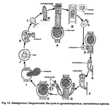
In Oedogonium the thallus is haploid and the life cycle is haplontic type. The diploid stage in life cycle is only zygote. It occurs for a short period. The zygote or oospore undergoes meiosis to make four meiozoospores which again form haploid Oedogonium thalli. The variations in life cycles of Oedogonium are due to macrandrous and nannandrous nature of Oedogonium species.
Macrandrous Forms:
Oedogonium macrandrous species can be monoecious or homothallic, if antheridia and oogonia are produced on same filament (Fig. 10, 11).
Oedogonium macrandrous species can be dioecious or heterothallic if antheridia are produced on male plants and oogonia are produced on separate female plants. (Fig. 12).
Nannandrous Forms:
The nannandrium or dwarf male plants are produced by germination of androspores which are produced in androsporangia. In gynandrosporous nannandrium forms the androsporangia and oogonia are formed on same filaments (Fig. 13, 14).
In idioandrosporous nannandrium forms, the androsporangia and oogonia are formed on different plants.


