ADVERTISEMENTS:
In this article we will discuss about Volvox. After reading this article we will learn about:- 1. Systematic Position of Volvox 2. Occurrence 3. Structure 4. Reproduction 5. Fertilization 6. Life cycle.
Systematic Position of Volvox:
Occurrence of Volvox:
Volvox is free floating fresh water green algae. Volvox grows as planktons on surface of water bodies like temporary and permanent ponds, lakes and water tanks. During rainy season due to its fast growth the surface of water bodies become green. The Volvox colonies appear as green rolling balls on surface of water.
ADVERTISEMENTS:
Volvox is represented by about 20 species:
Some common Indian species are—Volvox globator, V aureus, V. prolificus, V. africanus and V. rousseletii.
Structure of Volvox:
Volvox thallus is a motile colony with definite shape and number of cells. This habit of thallus is called coenobium.
ADVERTISEMENTS:
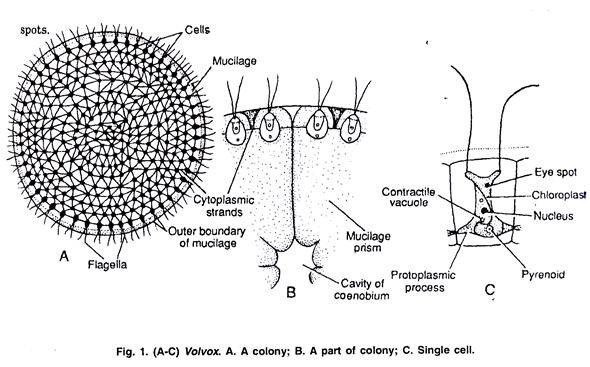
The colony is hollow, spherical or oval in shape and the size of colony is about the size of a pin head. The number of cells in a colony is fixed. Depending upon the species of Volvox the cells can be 500-60,000. The central part of colony is mucilaginous and the cells are arranged in a single layer on periphery of the colony (Fig. 1A).
The cells of anterior end possess bigger eye spots than those of posterior end cells. The cells of posterior side become reproductive on maturity. Thus, spherical or round colony of Volvox shows clear polarity. The cells of Volvox colony are Chlamydomonas type. Every cell has its own mucilage sheath (Fig. 1 B).
The mucilage envelope of colony appears angular due to compression between cells. The cells are connected to each other through cytoplasmic strands. In some species of Volvox the cytoplasmic connections or strands are not present.
The cells of colony are usually pyriform with narrow anterior end and broad posterior end. The cells are biflagellate, the two flagella are equal, whiplash type and project outwards (Fig. 1 C). The protoplasm of cell is enclosed within plasma membrane.
Each cell contains one nucleus, a cup shaped chloroplast with one or more pyrenoids, an eye spot and 2-6 contractile vacuoles. In some species of Volvox e.g., in V. globator and V. rousseletii the cells are of Sphaerella type.
The cells of colony are independent for functions like photosynthesis, respiration and excretion. The movement of colony takes place by co-ordinated flagellar movement. The reproduction is common to the coenobium.
Reproduction in Volvox:
Volvox reproduces both asexually and sexually. The asexual reproduction takes place under favourable conditions during spring and early summer. In Volvox mostly the cells of posterior part of colony take part in reproduction. These reproductive cells can be recognized by their larger size, prominent nuclei, dense granular cytoplasm, more pyrenoids and absence of flagella.
Asexual Reproduction:
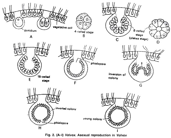
During asexual reproduction some cells of the posterior part of colony become reproductive. These cells enlarge up to ten times, become rounded and lose flagella. These cells are called gonidia (Sing, gonidium) (Fig. 2 A). The gonidia lose eye spot. Pyrenoids increase in number.
ADVERTISEMENTS:
The gonidia are pushed towards interior of the colony. The first division of gonidium is longitudinal to the plane of coenobium and this forms 2 cells (Fig. 2 A).
The second division is also longitudinal and at right angle to the first, forming 4 cells (Fig. 2 B). By third longitudinal division all the four cells divide to make 8 cells of which 4 cells are central and 4 are peripheral. These 8 cells are arranged in curved plate-like structure and are called plakea stage (Fig. 2 C, D). Each of these 8 cells divides by longitudinal division forming 16 cells arranged in the form of a hollow-sphere (Fig. 2 E).
The sphere is open on exterior side as a small aperture called phialopore (Fig. 2 F). The cells at this stage continue to divide till the number of cells reaches the characteristic of that species. The cells at this stage are naked and in close contact with each other. The pointed anterior end of cells is directed towards inside.
The next step is called inversion of colony (Fig. 2 G-H). As cells become opposite in direction, their anterior pointed end has to face the periphery of colony.
ADVERTISEMENTS:
The inversion of colony starts with formation of a constriction opposite to phialopore. The cells of posterior end along with constriction are pushed inside the sphere, till the whole structure comes out of the phialopore. After inversion, the anterior pointed end of the cell faces periphery.
The phialopore gets closed, and makes the anterior part of the colony. After inversion the cells develop cell wall, flagella and eye spot. The cells become separated due to development of gelatinous sheath around each cell. This newly developed colony is called daughter colony (Fig. 2 I).
The daughter colonies initially remain attached to gelatinized wall of parent colony and later become free in gelatinous matrix of parent colony. The daughter colonies are released in water after the disintegration of parent colony or through the pores. Sometimes next generation of daughter colonies develop while the colonies are still attached to the earlier parent colony.
Sexual Reproduction:
ADVERTISEMENTS:
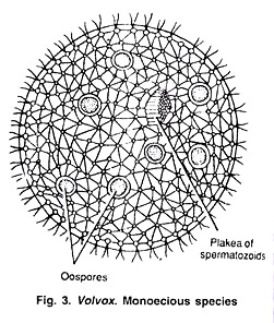
The sexual reproduction in Volvox is oogamous type. Some species of Volvox e.g., V. globator are monoecious or homothallic (Fig. 3) i.e., the antheridia and oogonia develop on same colony. Other Volvox species e.g., V. rousseletii are dioecious or heterothallic i.e., antheridia and oogonia develop on different colonies.
Monoecious species are usually protandrous i.e., antheridia mature before oogonia but some species are protogynous i.e., oogonia develop before antheridia. V aureus is mostly dioecious but sometimes can be monoecious.
Reproductive cells mostly differentiate in the posterior part of colony. These cells enlarge, lose flagella and are called gametangia. The male reproductive cells are called antheridia or androgonidia and female reproductive cells are called oogonia or gynogonidia.
Development of Antheridium:
ADVERTISEMENTS:
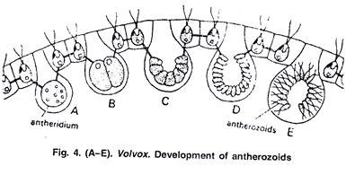
The development of antheridium starts with formation of antheridial initial or androgonidial cell mostly in posterior side of the colony. The initial cells enlarge, lose flagella, protoplasm becomes dense and nucleus becomes larger. The antheridial initial shifts inside towards cavity and remains connected to other vegetative cells through cytoplasmic strands.
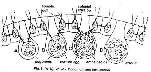
The protoplast of antheridial initial divides, longitudinally to form 16-512 elongated cells. The cells remain in plate like structure or arrange in a hollow sphere. The inversion of cells also takes place as in asexual reproduction. Each cell differentiates in antherozoid or spermatozoid (Fig. 3, 4).
The antherozoid is spindle shaped, elongated, bi-flagellated structure containing two contractile vacuoles, nucleus, cup shape chloroplast, pyrenoid and eye spot. It is pale yellow or green in colour. The antherozoids are released individually or sometimes in groups.
Development of Oogonium:
The oogonia also differentiate mostly in posterior side of the colony. The oogonial initials enlarge, nucleus becomes larger, protoplast becomes dense, flagella are lost, eye spot disappears and many pyrenoids appear. The mature oosphere or ovum is round or flask shaped structure. The egg is uninucleate structure, the beak of flask shape oogonium functions as receptive spot (Fig. 5 A, B).
Fertilization of Volvox:
After liberation from antheridium, the antherozoids swim freely on surface of water. Due to chemotactic response the antherozoids reach the oogonia.
Some antherozoids enter each oogonium. Only one antherozoid enters inside the oogonium through receptive spot. After this plasmogainy i.e., fusion of male and female cytoplasm and karyogamy i.e., fusion of male and female nuclei take place. This results in formation of diploid zygote (Fig. 5 D).
The diploid zygote secretes a three layered thick wall. The layers of the wall are exospore, mesospore and endospore (Fig. 6 A, B). The outer exospore is thick. It may be smooth e.g., V. aureus (Fig. 6 A) or spiny e.g., V. globator (Fig. 6 B).
The mesospores and endospores are thin and smooth. The walls contain nucleus pigment haematochrome which imparts red colour to the zygote. The zygotes are released by the disintegration of parent colony. Then zygotes undergo a period of dormancy.
Germination of Zygote:
The dormant zygote germinates on approach of favourable climatic conditions. The diploid nucleus of zygote undergoes meiotic division forming four haploid cells. The outer two layers of zygote burst and the inner layer comes out as vesicle. The four haploid cells migrate with the vesicle (Fig. 6 C, D). The development of new colony from zygote differs in different species of Volvox.
In V. aureus and V. minor the protoplasm of zygote divides repeatedly until the cell number of colony is reached and new colony is formed as in asexual reproduction process. In V. campensis the protoplast of zygote divides to make many biflagellate zoospores.
ADVERTISEMENTS:
Only one zoospore survives and all other disintegrate. This zoospore comes out of the vesicle it divides to make many cells which arrange to form a colony. In V. rousseletii the zygote forms a single biflagellate zoospore, the protoplast of zoospore divides and forms a colony. In all the methods the cells divide and undergo inversion to make a mature colony (Fig. 6 E-H).
Life Cycle of Volvox:
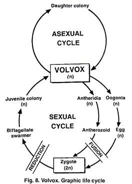
Volvox is haploid (n) algae, the haploid gametes fertilize to make diploid zygote (2n) which divides by meiosis to make haploid cells (n) which mature into haploid Volvox colony (Fig. 7, 8).

