ADVERTISEMENTS:
In this article we will discuss about:- 1. Occurrence of Chlamydomonas 2. Plant Body of Chlamydomonas 3. Features 4. Reproduction 5. Life History.
Occurrence of Chlamydomonas:
The genus Chlamydomonas (Gr. Chlamys, mantle; monas, single organism) includes about 500 species, found almost everywhere (i.e., ubiquitous). Commonly they are found in fresh water of lakes, ponds, tanks etc., but they are also available in brackish water (C. halophila), saline water (C. ehrenbergii), snow (C. nivalis) and some are also air borne.
The snow of arctic and alpine zones becomes red due to the presence of C. nivalis which accumulates the red pigment haematochrome. But the snow covered mountain range of yellow stone national park (USA) becomes yellowish green due to the well- developed population of C. yellowstonensis.
Plant Body of of Chlamydomonas:
ADVERTISEMENTS:
Plant body of Chlamydomonas is unicellular and motile (Fig. 3.41). The cells are usually spherical, oval or oblong in shape but other forms like ellipsoidal, pyriform etc. are also available (Fig. 3.39). Most of the species are broader towards the posterior side and pointed towards the anterior side, which gradually ends in apical papilla.
The length of the cell varies from 20 to 30μm but rarely exceeds 30μm in major diameter.
The cell wall is thin, smooth and firm. It is made up of cellulose. The major structural component of cell wall is glycoprotein. In some species (C. gleocystiformis), the cell wall is surrounded by mucilaginous pectin layer formed by the dissolution of pectose layer to pectin.
ADVERTISEMENTS:
Inner to the cell wall, semipermeable cell membrane or plasma membrane is present which surrounds the protoplast.
The protoplast contains the following:
a. Chloroplasts:
It occupies the lower broader part. It is generally of cup-shaped and parietal (Fig. 3.40). (However, the chloroplast is variable in shape, such as H-shaped in C. biciliata; parietal in C. mucicola; reticulate in C. reticulata and stellate in C. arachne).
Chloroplast has single pyrenoid with a starch sheath. The number is also variable and it may be two (C. debaryana) to many (C. gigantae). Sometimes they may be numerous and distributed irregularly inside the chloroplast (C. sphgnicola).
b. Eye-Spot:
Towards the anterior end of the chloroplast at one side an circular to oval, photoreceptive organ, the stigma or eye spot is present. The spot consists of a curved pigmented plate,, the pigmentosa and a biconvex lens.
c. Nucleus:
The nucleus remains suspended inside the cups and is of prokaryotic in nature.
ADVERTISEMENTS:
d. Other Inclusion:
It includes mitochondria, E.R. vesicles, contractile vacuoles etc.
e. Flagella:
Two whiplash-types of flagella are present towards the anterior region of the cell. They are equal in length. The flagella may be very small, same size of the cell or bigger than the cell in most species.
ADVERTISEMENTS:
The flagellum originates from the blepharoplast, situated towards the anterior side (Fig. 3.42). The flagella come out through very fine canals, on the outer wall.
In some species like C. nasuta neuromotor apparatus (Fig. 3.42) is said to be present. The neuromotor apparatus consists of two basal granules, the blepharoplasts. Both are connected by a fibre, called paradesmose. One of the blepharoplast is connected to the centrosome of the nucleus by a thread, the rhizoplast. Many fine fibrils connect the centrosome with the nucleolus.
The E.M. studies have not confirmed about the neuromotor apparatus:
f. Contractile Vacuoles:
ADVERTISEMENTS:
Two contractile vacuoles are present just below the blepharoplast. Possibly they regulate the water content of the cell, by discharging more water at times.
Structure as observed under E.M.:
Roberts et al (1972), Hills (1973) and Hills et al. (1973) have studied in detail the structure of Chlamydomonas under Electron Microscope (E.M.) (Fig. 3.43).
The cell wall is multilayered (7 layered) and it consists of proteins. Cellulose is absent. Cell membrane like normal eukaryotic cell is lipoprotein in nature. The flagellum remains attached to the basal granule, the blepharoplast and shows typical 9 + 2 fibrillar arrangement. The chloroplast is cup-shaped (C. eugametor) and surrounded by double-layered unit membrane.
It bears a number of band-shaped photosynthetic lamellae, the thylakoids; are lipoprotein in nature and remain dispersed in the granular matrix, the stroma. The grana-like bodies are formed by the aggregation of about 3-7 thylakoids. The chloroplast matrix contains ribosomes, microtubules, crystal-like bodies etc.
ADVERTISEMENTS:
The cytoplasm contain nucleus, mitochondria, Golgi apparatus, E.R, ribosomes etc. The nucleus always lies in the cytoplasm, present in the cavity of chloroplast.
The eye-spot consist of 2-3 parallel rows of lipid droplets (i.e., granules) and each one measures about 75nm in diameter. It remains embedded at one side of the chloroplast.
Important Features of Chlamydomonas:
1. Plant body is unicellular, pear-shaped and biflagellate.
2. Each cell has generally cup-shaped chloroplast, one eye-spot and two contractile vacuoles.
3. Presence of palmella-stage.
4. Asexual reproduction takes place through biflagellate zoospore formation.
ADVERTISEMENTS:
5. Sexual reproduction through iso-, aniso-, and oogamy.
Reproduction in Chlamydomonas:
Chlamydomonas reproduces both asexually and sexually:
Asexual Reproduction:
Asexual reproduction takes place commonly by the zoospore formation. But some others also reproduce by aplanospores, hypnospores, palmella stage and rarely by synzoospores.
1. Zoospores:
During night with favourable environmental conditions zoospores are formed (Fig. 3.44). At the starting of zoospore formation, the parent cell withdraws its flagella and takes rest. The contractile vacuoles disappear and the protoplast slightly withdraws from the cell wall. The protoplast then undergoes repeated longitudinal division at right angles to one another and forms generally 8 but rarely up to 64 units.
Each unit of protoplast secretes a wall around and develops contractile vacuoles. They are released by rupturing or gelatinisation of the mother wall. At the time of liberations they develop their flagella. These flagellated daughter units are called zoospores. After liberation they behave as new individuals and capable of developing new crop of zoospores after 24 hours.
2. Aplanospores:
The aplanospores are formed during unfavourable conditions. Following the same procedure like zoospore-formation, they develop 2-16 daughter protoplasts. Each one secretes, thin wall around, itself. These thin walled non-motile spores are called aplanospores. During favourable conditions aplanospore comes out of the mother cell and develops a new cell directly or its protoplast divides to form more zoospores.
3. Hypnospores:
The hypnospores are formed during severe drought. The hypnospores are developed like aplanospores, but the wall becomes much thicker than aplanospores. During favourable condition they germinate like aplanospores i.e., either directly or by the formation of zoospores through more divisions of protoplasts.
4. Palmella Stage:
During zoospore formation, suddenly if the environmental condition becomes unfavourable, the parent wall gets gelatinised. Consequently the daughter protoplasts also get gelatinised and undergo divisions. The entire structure becomes enlarged much more. This stage looks like another green alga Palmella of Tetrasporales and called this stage as Palmella stage (Fig. 3.45).
During favourable conditions, the unit bodies develop into individual zoospore. Palmella stage is very common in C. kleinii and C. braunii.
5. Synzoospore:
The multinucleate and multi- flagellate zoospores are called synzoo- spores. They are reported to be formed in artificial culture. The mother nucleus divides into 4 nuclei, those develop a pair of flagella and finally to synzoospores.
Sexual Reproduction:
Sexual reproduction takes place during desiccation or deficiency of nitrogenous compound in the growing medium. It takes place very commonly through isogamy and less frequently through anisogamy and oogamy.
1. Isogamy:
Majority of the species are isogamous. In this type sexual union takes place between the morphologically identical gametes (Fig. 3.46).
In some species the vegetative cell directly develops into gametes (hologamy) or generally it divides into 8 to 64 gametes. The uniting gametes may develop from the same plant (e.g., C. debaryanum) i.e., homothallic or from different plants i.e., heterothallic (e.g., C. reinhardii and C. moewusii).
During union -the flagella are found to be covered by agglutin (not found on the flagella of vegetative cell), a chemical substance (protein) which helps in the recognition of gametes of opposite mating type.
In most of the species the gametes are unicellular, uninucleate, very small and biflage- llate structures (Fig. 3.46A).
During sexual union isogametes (+ and -) come very close to each other. The wall at the point of contact dissolves resulting in the formation of diploid quadriflagellate zygote through plasmogamy followed by karyogamy (Fig. 3.46B-D).
2. Anisogamy:
It is the union between two gametes (male and female) of different sizes (Fig. 3.47A, B). The larger one is called macrogamete (female) produced only 2 or 4 in the female gametangium. The smaller one is called microgamete (male) produced 8 or 16 in the male gametangium.
The micro- gametes are more active than macrogametes. The microgamete comes very close to the macrogamete. Both the gametes undergo fusion and form zygote (Fig. 3.47C-E). This type of sexual union is found in C. braunii, C. suboogama etc.
3. Oogamy:
It is the union between morphologically dissimilar gametes (Fig. 3.48A, B). The microgamete (male) is biflagellate and smaller in size than the macrogamete (female) which is non-motile and larger .n size. The male cell divides repeatedly and forms 16 units, each of which is converted into male gamete.
On the other hand, the female cell leaves the flagella and directly functions as female gamete. The active male gamete comes very close to non-motile female gamete and attaches itself at the anterior end. Both the gametes undergo fusion and form zygote (Fig. 3.48C-D). This type of union is found in C. coccifera.
Zygote:
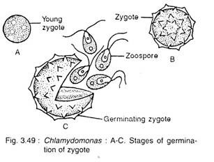
Initially the quadriflagellate zygote remains motile for several hours (C. paradoxa) to about 15 days (G pertusa). After leaving flagella it settles down on the substratum and takes rest. Primary wall is formed by the zygote, followed by thick ornamented secondary wall (Fig. 3.49A, B). With maturity the zygote accumulates large amount of starch and oil.
Germination of Zygote:
During germination (Fig. 3.49C) the diploid nucleus -(2n) of the zygote undergoes meiosis to form 4 (G chlamy- dogama) or with additional mitosis forms 16 to 32 (G intermedia) nuclei (n). The segregation of + and – strains takes place during meiosis. The inner wall dissolves and by breaking the outer wall the haploid cells are liberated. During liberation they develop flagella and behave like new individuals.
Indian Species:
Chlamydomonas eugametos, C. grandistigma, C. ehrenbergii.
Life History of Chlamydomonas:
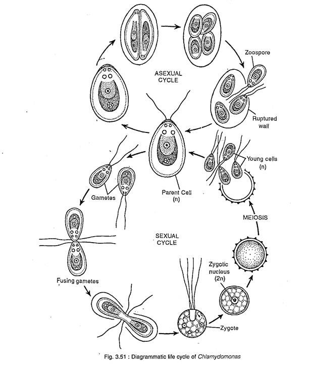
Life history of Chlamydomonas is given in Fig. 3.50 and 3.51:


