ADVERTISEMENTS:
In this article we will discuss about the cell structure of cyanophyceae with the help of diagrams.
The cyanophycean cells are prokaryotic in nature, which rarely exceeds 10p in diameter. Each cell consists of outer covering of cell envelop which surrounds the membrane covered protoplast (Fig. 3.24C).
1. Envelop:
ADVERTISEMENTS:
It consists of outer mucilaginous sheath and inner cell wall.
a. Mucilagenous Sheath:
Presence of mucilaginous sheath is common in all cyanophycean members. It consists of three layers of microfibrils arranged reticulately within an amorphous matrix.
ADVERTISEMENTS:
This layer endows the cells with greater water absorbing and retaining capacity which helps to survive during desiccation and forms a barrier for parasites.
b. Cell Wall:
The cell wall consists of four (4) (Fig. 3.24A) layers (under E.M.) named as LI, LII, LIII and LIV by Carr and Whitton (1973). Each layer is about 10µ in thickness. The LI is the layer situated near cell membrane and LIV is the outermost.
Cell wall is composed of mucopeptide together with carbohydrates, amino acids and fatty acids like Gram-positive bacteria. The LI and LIII layers are electron transparent, but the LII and LIV layers are electron opaque (impervious).
2. Cytoplasmic Membrane:
The cytoplasmic membrane is also known as plasmalemma present just inner to the cell wall. It consists of two electron opaque layers separated by a translucent layer (Fig. 3.24A).
This layer invaginates at different points inside the protoplast and is the site of different biochemical functions that normally takes place in different organelle like mitochondria, endoplasmic reticulum and Golgi bodies in the cells of eukaryote.
3. Protoplast:
Studies with Electron Microscope by Wilden and Mercer, Pankrats and Bowen and many others show that the protoplast consists of thylakoids, cytoplasmic inclusion and nucleoplasm.
a. Thylakoids:
These are the complex lamellar system, which functions like the protoplasts of eukaryotes. Thylakoids are not bounded by membrane instead they appear as elongated and flattened sacs composed of two unit membranes.
ADVERTISEMENTS:
Each membrane is about 75Å thick. The neighbouring thylakoids are separated by a flattened space of 50nm. The space is occupied by rows of discoid phycobilisomes, which contain the photosynthetic pigments chlorophyll a, c-phycocyanin and c-phycoerythrin.
b. Cytoplasmic Inclusions:
The cytoplasmic inclusions present in the cyanophycean cell are ribosomes, cyanophycean granules, polyhedral bodies, polyphosphate bodies, polyglucoside bodies, α-granules, β-granules and gas vacuoles (Fig. 3.24C).
The function of the above inclusions is not well-known. Cyanophycean granules are regarded as reserve food, polyhedral bodies, i.e., the carboxysomes contain enzyme ribulose b is phosphate carboxylase oxygenase.
ADVERTISEMENTS:
Ribosomes take part in protein synthesis. Some workers think α-granule as reserve food, a glycogen-like substance. Many prokaryotic members contain many small gas vacuoles also called pseudo- vacuoles. They help to float in water. They refract light and the cells become brown, purplish or black under microscope.
c. Nucleoplasm:
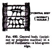
The nucleoplasm is usually centrally located and contains numerous fine randomly oriented fibres of DNA. It is differentiated from the eukaryotic nucleus in absence of nucleolus and nuclear membrane. They are not differentiated from the cytoplasm by any membrane and are present in close association (Fig. 3.24C).
The region is with low electron density than the surrounding cytoplasm. The DNA does not organise into chromosomes due to the absence of protein like histones or protamines.
4. Heterocysts:
ADVERTISEMENTS:
These are specialized cells of the filament distinguished from others by their thick wall, polar nodule(s) and homogenous contents.
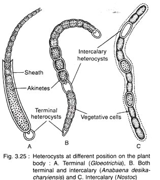
Heterocysts are commonly found in the members of Stigonematales and Nostocales (except Oscillatoriaceae). They grow in the filament either terminally (Gloeotrichia) or intercalary (Nostoc) or both terminal and intercalary (Anabaena desikacharyiensis) (Fig. 3.25).
Structure:
ADVERTISEMENTS:
Pale yellow, thick-walled specialized cells, larger than vegetative cells are called heterocysts. The wall of the heterocyst is differentiated into three regions, an outer fibrous, middle homogenous and inner lamellar layer (Fig. 3.26).
It has pore(s) at the pole(s) where it remains attached with the vegetative cell. The pores are plugged with polar nodule(s) or polar granules. The wall of the heterocyst is thicker towards the polar regions.
The inner content is dense and appears to be homogeneous. The thylakoides i.e., the photosynthetic lamellae are present, but the ribosomes are less in number. Other granular inclusions are appears to be absent. Due to absence of phycobillins and chlorophyll a, photosynthesis does not take place.
The thylakoids are tightly packed due to reduction of interlamellar space and are present more towards the periphery than the central region. Due to low concentration or absence of pigment, the mature heterocyst appears to be empty. The two Iipids, glycolipid and acyllipid present in the lamellae are not found in the vegetative cells.
Development of Heterocyst:
ADVERTISEMENTS:
It develops either singly or in pair from single or both the daughter cells of a recently divided vegetative cell. The daughter cell may behave as proheterocyst, which later on develop into a heterocyst.
The developmental changes are described below:
i. The inner content of protoplasm becomes paler in colour.
ii. The wall of the polar region becomes rounded.
iii. The connecting region (s) between vegetative cell and heterocyst becomes more constricted.
iv. Formation of an inner polysaccharide wall external to the cell membrane.
ADVERTISEMENTS:
v. Formation of pores at one (terminal) or both (intercalary) ends.
vi. Protoplasmic connection being established with the adjacent vegetative cell(s) through the pores.
vii. The pores become plugged by mucilage towards maturity, which are called nodule.
viii. The content of the cell becomes homogeneous with different chemical changes.
ix. Gradual breakdown of thylakoids and dissolution of storage granules.
ADVERTISEMENTS:
Function of Heterocyst:
The function of heterocyst is not clearly known.
Many scientists have put forth different suggestions regarding the function of heterocyst:
1. Some considered it as the store-house of food materials.
2. Some others considered that they help in hormogonia formation.
3. According to Brand (1903), it is of spore-like structure.
4. Smith (1951) pointed out it as vestigial spore.
5. Fritsch (1951) told that during vegetative period it secretes certain substances which promote growth and cell division.
6. According to Fay (1968) it is the site of N2 fixation.
7. Singh and Kumar (1970) pointed out that in addition to N2 fixation; it also helps in growth and development of the filament.
8. Later, Steward (1970) suggested the possible roles of heterocyst such as:
i. Involvement in N2 metabolism,
ii. Growth and differentiation,
iii. Control of sporulation, and
iv. Antiquated reproduction unit.


