ADVERTISEMENTS:
Read this article to learn about the Reproductive System and Life Cycle in Ascaris !
Systematic Position
Phylum: Aschelminthes
Class: Nematoda
ADVERTISEMENTS:
Order: Ascaroidea
Genus: Ascaris
Species: lumbricoides
Ascaris lumbricoides is elongated, cylindrical, and tapering at both ends. It is a large sized nematode showing sexual dimorphism, i.e., sexes are separate. They can be easily distinguished externally, i. e the male is smaller in size than the female, in male tail is curved.
 The anterior end of the body bears a terminal, triradiate mouth which is surrounded by 3 crescentic lips- one median dorsal and two ventro-laterals. Each lip bears minute teeth-like denticles along its inner, oral border and small sensory outgrowths. There is a pair of minute cervical sensory papillae on the sides of body, and a small excretory pore in the midventral line. A pair of minute lateral glandular receptors, phasmids is present in the form of distinct cuticular notches or pits.
In females Ascaris, there is the genital pore or vulva; males do not have a separate genital pore. The anus in these serves for genital pore also and, it is called cloacal aperture. Two small needle-like penial spicules or copulatory seatae, formed of cuticle, protrude out from the cloacal aperture (fig. 9.16 A, B).
Only sexual reproduction occurs in Ascaris. Sexes are separate and there is distinct sexual dimorphism between male and female Ascaris.
Male Reproductive System:
Reproductive organs of male ascaris include a testis, a vas deferens, a seminal vesicle, an ejaculatory duct, cloaca and penial setae.
1. Testes:
Male ascaris is monorchis i.e., it has a single testis. The testes are a long, thin and coiled tube-like structure. It continues into a vas deferens. The testis has a cavity lined by a single layer of cuboidal cells. It acts as “growth zone”. It has, in its cavity, a semisolid axial core of rachis of cytoplasm around which the spermatogonia are irregularly attached while these progressively undergo maturation or spermatogenesis to form sperms.
2. Vas deferens:
This is shorter and less coiled tube than testis.
ADVERTISEMENTS:
3. Seminal vesicle:
This is a long, straight and relatively much thicker tube into which the vas deferens opens. It serves to store the mature sperms.
4. Ejaculatory duct:
The seminal vesicle leads into a short, narrow and highly muscular and glandular ejaculatory duct. This duct opens behind into the last part of rectum from the ventral side.
ADVERTISEMENTS:
5. Cloaca:
The last part of rectum, located behind the opening of ejaculatory duct serves as cloaca, because it receives both faeces and sperms. It opens out by the cloacal aperture.
6. Penial setae:
Two small, contractile penial sacs open into the cloaca on dorsal side. Each sac secretes and contains a small needle-like penial or copulatory seta or spicule of cuticle. Protractor and retractor muscles, associated with the wall of each penial sac respectively serve to protrude and retract the contained spicule through the cloacal aperture. During copulation, the spicules protrude out to keep the female’s vulva open. Sperms of Ascaris are tail, asymmetrical and amoeboid.
ADVERTISEMENTS:
Female Reproductive System:
The female genitalia include ovaries, oviducts, uterus and vagina.
1. Ovaries:
Female Ascaris is didelphic i.e., it has two ovaries. It is long, thin and coiled tube-like structures. The oogonia are formed by budding from a single large germinal cell forming the proximal “germinal zone” of each ovary. In the remaining part of an ovary, i.e., the “Growth zone”, oogonia undergo oogenesis or maturation, The oogonia after completing the first maturation divisions, become secondary oocytes and reach the oviducts.
ADVERTISEMENTS:
2. Oviducts:
Each ovary leads into a long and coiled oviduct.
3. Uterus:
Each oviduct leads into a much thicker and long uterus. The uterine wall thick and formed of a layer of tufted secretory cells surrounded by a muscular layer Uteri serve to store the eggs after fertilization.
4. Vagina:
The uteri open into short and relatively narrow vagina. The wall of vagina is quite muscular and contractile. The vagina opens out by slit-like female pore or vulva.
ADVERTISEMENTS:
Life Cycle:
Copulation and fertilization:
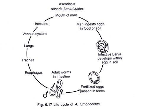
Ascaris copulates in the intestine of the host. The sperms by amoeboid movement reach the seminal receptacle of the uterus and fertilize the ovum. Fertilized eggs pass one-by-one into the uteri. A sphincter muscle regulates their passage from the seminal receptacle into the remaining part of a uterus, (fig. 9.17).
Mammilated Eggs:
Immediately after fertilization, the cell membrane of a zygote separates from its cytoplasm and, thus, becomes a fertilization membrane. Soon, it is fortified by protein and glycogen granules extruded by the cytoplasm. Thus, it transforms into a thick and hard chitinoid shell.
As the eggs move to the uteri, the cells of uterine wall secrete a brownish albuminous cyst around each egg. On drying, this cyst becomes hard, rough and warty. Thus, by the time an egg reaches the distal part of a uterus, it becomes enclosed in three, highly resistant protective coverings and bears distinct warts or tubercle upon its surface. It is called a mammilated egg.
ADVERTISEMENTS:
Cleavage and early embryonic development:
Embryonic development is possible in Ascaris only outside the body of human host in soil, because it requires low temperature, more O2 and suitable moisture. Cleavage is holoblastic, but of a peculiar spiral and deteminate type. In quite early stages, different blastomeres of the embryo become earmarked to form different organs of future juvenile ascaris.
Infection of host:
Ascaris has a single-host life cycle i.e. monogenetic life cycle. Eggs containing the second stage juvenile are called embryonated eggs. These are infective to human host. People acquire infection by ingesting embryonated eggs with contaminated food and water.
When these eggs reach into the intestine of human host, their coverings are digested and the juveniles, measuring become free in intestinal lumen. Boring through the intestinal wall, these juveniles make their way into the sub-mucosal blood capillaries. Then, with the blood circulation, these undergo an extensive migration in the host body.
This migration is divisible into two phases:
(i) Primary migration:
During this migration, the juveniles remain in blood of host for about a week. From the capillaries of intestinal wall, these reach into liver in about 3 to 4 days via the hepatic portal system. Then, in another 3or 4 days, these reach into the lungs via the hepatic veins, heart and pulmonary arteries.
(ii) Secondary migration:
This migration takes place in respiratory organs and alimentary canal of the host. In lungs the juveniles make their way out from the blood capillaries into the alveoli. Here, they grow in size, and moult their cuticle twice, to allow the growth of body. Thus, the second stage juveniles change into third and fourth stage juveniles in lung alveoli.
The fourth stage juveniles start ascending up the bronchi and trachea of the host who, therefore, suffers from coughing for this reason. Hence, the juveniles are coughed into the pharynx of the host and are swallowed into oesophagus. Shortly thereafter, these travel through stomach and reach back into the intestine.
After reaching back into the intestine, the juveniles soon undergo fourth and the last moult of their cuticle and become adults. Within 8 to 10 weeks, adult ascaris starts reproducing. Its life span in the host is of 9 to 12 months. After this, it dies and disintegrates.
Aberrant migration:
Sometimes, the juveniles migrate into brain, spinal cord, eyes etc., with blood from the heart, instead of migrating into the lungs. In such organs, these soon become dead and encysted in calcified cysts. Similarly, during their migration from trachea into the pharynx, strong coughing may cause ejection of juveniles through nose or mouth of the host.
Ascaris infection can be prevented by maintaining cleanliness and good sanitary conditions. Vegetables should be thoroughly washed (preferably in a mild solution of KMnO4) and properly cooked before use. Washing the hands with antiseptic soap before touching edible materials may help reduce the infection of ascaris.

