ADVERTISEMENTS:
In this article we will discuss about Subphylum Vertebrate (Craniata):- 1. Introduction to Subphylum Vertebrate (Craniata) 2. Characters of Subphylum Vertebrata (Craniata) 3. Basic Features 4. Origin 5. Ancestry.
Contents:
- Introduction to Subphylum Vertebrate (Craniata)
- Characters of Subphylum Vertebrata (Craniata)
- Basic Features of Subphylum Vertebrata (Craniata)
- Origin of Vertebrates
- Ancestry of Vertebrates
1. Introduction to Subphylum Vertebrate (Craniata):
ADVERTISEMENTS:
The vertebrates constitute the main subdivision of the Phylum Chordata and occupy the highest rank. All the vertebrates or craniates are placed under the Subphylum Vertebrata or Craniata.
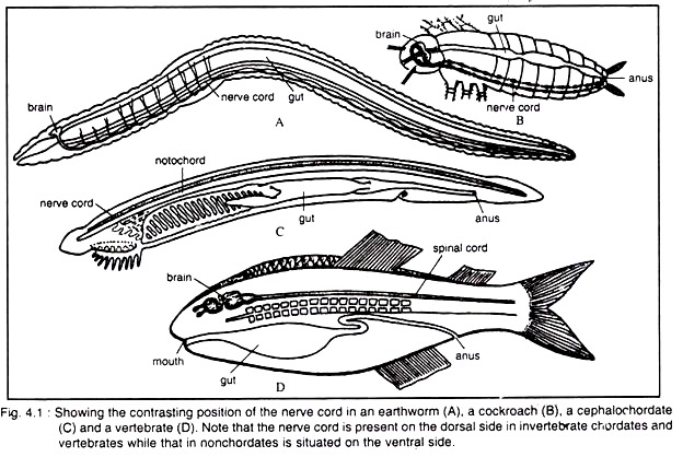
In addition to the basic chordate features, the vertebrates possess many specialised characteristics that distinguish them from the invertebrate chordates and non-chordates. The main difference lies in the position of the nerve cord (Fig. 4.1).
Subphylum Vertebrata (Craniata):
ADVERTISEMENTS:
The vertebrates constitute the main subdivision of the Phylum Chordata. All the vertebrates or craniates are placed under the subphylum Vertebrata or Craniata. With the new information to the Zoologists, they are inclined to create more subphyla rather than Vertebrata.
Nelson (1994) has mentioned in his scheme the subphyla Conodontophorida and Craniata, other than Vertebrata. In general sense the Vertebrata or Craniata is used in most recent literature.
2. Characters of Subphylum Vertebrata (Craniata):
a. All vertebrates have endoskeleton framework.
b. All the members possess cranium but vertebrae are usually.
c. Due to the presence of vertebral column, the group derives its name from this structure.
d. The notochord does not extend beyond the brain.
e. The distinct cranium or braincase houses the brain. It is a major innovation of the vertebrates. The cranium bears also sensory capsules.
f. All vertebrates possess a well-developed head, i.e., cephalization is well-marked.
ADVERTISEMENTS:
g. The anterior part of the nerve tube becomes specialised to form a complex structure, called brain. The basic organisation of brain is similar in different vertebrates, but variations are due to regional specialisation of different parts, especially the cerebral hemispheres.
h. 10 to 12 pairs of cranial nerves.
i. Dorsal and ventral roots are usually united but in lampreys the dorsal and ventral roots remain separate.
j. Major part of the nervous system develops from ‘neural crest cells’, the embryonic cells that are found only in vertebrates.
ADVERTISEMENTS:
k. The heart is chambered. Although the heart in different classes exhibits diversity but the structure is basically similar in the different groups.
l. Distinct blood vessels and red blood corpuscles are present.
m. Hepatic portal system is present in all vertebrates.
n. The excretory organs are kidneys, which are of mesodermal origin that regulate the osmotic pressure and also excrete the nitrogenous wastes.
ADVERTISEMENTS:
o. Endostyle is present in ammocoete larva of lampreys and it transforms into thyroid gland in all other vertebrates.
Remark:
Most authors are in favour of using the terms — Vertebrata or Craniata, as synonyms. But Janvier (1981) and Nelson (1994) use the two terms for different levels of classification.
Janvier places all the groups of Vertebrates under Craniata, and Myxini is excluded from the Vertebrata, because the group (Myxini) lacks arcualia which are the embryonic or rudimentary vertebral elements. Parker (1982) uses the taxon subphylum Craniata instead of Vertebrata.
ADVERTISEMENTS:
3. Basic Features of Subphylum Vertebrata (Craniata):
ADVERTISEMENTS:
The subphylum Vertebrata or Craniata includes all craniates ranging from the agnathans to mammals. The body of vertebrates is built on a basic fundamental plan with modifications for living in different conditions. For better understanding a detailed discussion on two basic vertebrate characteristics like the vertebral column and the skull has been made in the following lines.
Vertebral Column:
All vertebrates have at least traces of vertebral column which forms the supporting and protecting axis of the body. It also offers sites for muscle attachment for locomotion, and gives rigidity to a soft body. In some aquatic forms integumentary respiration, thermoregulation and reception of external stimuli are performed to a certain degrees by an internal skeleton, specially the vertebral column.
In almost all the vertebrates, the notochord is replaced by the vertebral column in adult.
Development:
ADVERTISEMENTS:
During development, the mesodermal cells (Sclerotome) around the notochordal sheath become aggregated to form the skeletogenous layer (Fig. 4.2A). Some mesodermal cells also migrate into the notochordal sheath. By this way the notochord becomes surrounded by an investment of mesodermal cells. This- cellular investment transforms into cartilage which is called perichordal tube.
The skeletogenous layer then gives origin to the neural arch which takes the appearance of an inverted funnel, called cerebrospinal cavity. The skeletogenous layer also gives origin to paired haemal ridges which, in the posterior region of many vertebrates, unite to form the haemal canal. The developing vertebral column is broken up into segments which thus form a chain of bones, called vertebrae.
Parts of a typical vertebra:
A typical vertebra consists of a main body, the centrum bearing one or two arches and one or more attaching processes. The centrum develops as a ring of tissue around the notochord. The ring then fills in to become a disc or cylindrical mass. On the dorsal side of the centrum a neural arch becomes fused to enclose a neural canal through which the spinal cord passes.
A neural spine usually is developed from the mid-dorsal part of the neural arch. A similar arch is present in the caudal region of many vertebrates on the ventral side of the centrum. This ventral arch is called the haemal arch with haemal canal and haemal spine. Through the haemal canal the caudal artery and vein run (Fig. 4.2B).
ADVERTISEMENTS:
Besides these structures, there are several outgrowths or processes arising from the centrum and at the basal regions of the neural arch, called apophyses. These processes are projected in different directions and offer surfaces for attachment of muscles or facilitate frictional contact of one vertebra upon others as sliding joints.
The most common apophyses (Fig. 4.2B) are:
(a) Zygapophyses:
On the arch there are usually two pairs of Zygapophyses, articulate between successive vertebrae. The pair of zygapophyses projecting anteriorly are called prezygapophyses while the posterior pair, projecting at the basal region of the neural arch, called postzygapophyses. Prezygapophyses of the centrum face upward and postzygapophyses face downward. Zygapophyses are not common among fishes.
(b) Basapophyses:
Basapophyses are a pair of ventral projections of the centrum which represent the remains of the haemal arch.
(c) Diapophyses:
A pair of lateral projections of the centrum, called diapophyses. The diapophyses provide articulating surfaces for the dorsal head of the ribs, the tuberculum.
(d) Parapophyses:
A pair of lateral projections of the centrum which are used for the attachment of the ventral head of the ribs, the capitulum.
(e) Transverse process:
Laterally extended process which arises from the sides of the vertebra. Transverse processes are common among tetrapods.
(f) Hypapophyses:
Hypapophyses are the ventral projections of the centrum which are found in reptiles, birds and mammals.
Types of Vertebrae in living vertebrates:
The vertebrae constituting the vertebral column remain articulated end to end. The ends of the vertebrae vary in shape as a result the joint between the centra differs in different vertebrates, even in different regions of the vertebral column of a vertebrate.
Flower (1885), Weichert (1951), Walter and Sayles (1965), Hildebrand (1982), Romer and Parsons (1986), Kent and Miller (1997) and Kardong (1998, 2002) have mentioned the following types of vertebrae in their literature.
They are:
(a) Amphicoelous,
(b) Procoelous,
(c) Opisthocoelous,
(d) Heterocoelous and
(e) Amphiplatyan or acoelous (Fig. 4.2 C-H).
Amphicoelous vertebra:
(Gk. amphi = both, Gk. coel = cavity)
When both the ends of centrum are concave, the vertebrae are called amphicoelous. The centra touch each other at the rim of the intervertebral joint. The notochord passes through the aperture of the centrum and the concavities are filled with connective tissue and cartilage.
The spinal cord runs through the neural canal. Limited flexion of the body is affected in this type of joint. This type of vertebra is found in most fishes, primitive amphibians (stegocephalians), some living amphibians (e.g., Necturus, Proteus), primitive reptiles (cotylosaurs) and living reptiles like Spenodon, Gecko and in some reptilian forms.
Procoelous vertebra:
[Gk. pro = anterior, Gk. coel = concavity]
When the anterior end of centrum exhibits concavity and the posterior end shows convexity, the vertebrae are called procoelous. The procoelous vertebrae are found in some anurans (except sacral region of Bufo and Rana) and modern reptiles with a few exceptions. In the procoelous vertebra, the concavity is shallow in anurans but deep in reptiles.
Opisthocoelous vertebra:
(Gk. opisthe = behind, Gk. coel = concavity)
When the centrum bears concavity at the posterior surface of the vertebra but the anterior surface shows convexity, a condition just reverse to the procoelous condition, is called opisthocoelous type vertebra.
This type of vertebra is found in Lepidosteus (a primitive of bony fishes), some anurans (Discoglossidae, Pipidae, etc.), 2nd and 3rd cervicals of turtles, penguin and parrots. The cervical vertebrae of ungulates are, and the cervical vertebrae of dinosaurs were opisthocoelous.
In procoelous and opisthocoelous, the convex articular surface of one vertebra fits with the concave articular surface of another vertebra, thus acts a ball and socket joint permitting a greater flexion in all directions of the body.
Heterocoelous vertebra:
(Gk. hetero = different, Gk. coel = concavity)
When the centrum of the anterior side of vertebra is convex dorsoventrally and concave from side to side, and the posterior face of the centrum is just reverse (concave dorsovenrally and convex from side to side), called heterocoelous vertebra, that is having a transverse, saddle-shaped surface in front and also saddle-shaped surface behind vertically.
Great freedom is the lateral and vertical flexion of the neck region but prevents in the rotation of the vertebral column, e.g., cervical vertebrae of birds.
Amphiplatyan vertebra or Acoelous:
(Gk. amphi = both, Gk. platys = flat)
If the centrum of the vertebra is flat at both sides, it is called amphiplatyan, also known as acoelous. This type of articular surfaces helps to receive and distribute compressive forces within the vertebral column. These vertebrae are characteristics of mammals.
In some eels the anterior and posterior surfaces of the centra are flat. Infrequent types of vertebrae are seen in the vertebrates such as biconvex type (e.g., 9th vertebra of Bufo and Rana, 4th cervical of turtles), platycoelous meaning flat in front and concave behind, e.g., in some mammals, and coeloplatyan that is concave in front and flat behind, e.g., in some mammals.
Jollie (1962) mentions the term ginglymoidy in cases of double articular surfaces of the vertebra such as double convex, concave or asymmetrical.
Phylogeny:
Evolution of the vertebral column is not entirely clear, especially during its phylogenetic inception. The earliest vertebrates, such as fossils of Myllokunmingia and Haikouichthys, and the living hagfishes possess a notochord but lack vertebrae. In lamprays a few small cartilaginous elements are seen, but vertebrae are absent.
In ostracoderms, we find a hint of vertebral column. Since then, the evolution of vertebral column in fishes and tetrapods is most complicated, because some parts of vertebral column are enlarged, others were lost, and some were evolved independently several times.
Skull:
The skull or cephalic skeleton in vertebrates is a double structure—both embryo- logically as well as morphologically. During embryonic development two sets of bones of different origin join together in a unified way.
The vertebrate skull is derived morphologically from two sources: the neurocranium (surrounding the anterior end of the nerve tube) and the splanchnocranium (encircling the anterior end of the digestive tube). The skull also plays double role—support and protection.
The skull starts its origin as paired cartilaginous plates (parachordals) situated one on either side of the notochord (Fig. 4.3A). Another pair of cartilaginous rods (trabeculae) develop in front of the parachordals.
Simultaneously, cartilaginous investments start formation around three paired special sense organs—olfactory capsules around the organs of smell, auditory capsules around the ears and the optic capsules around the eyes.
The olfactory capsules unite with the trabeculae, the auditory capsules unite with the parachordals and the optic capsules remain free. In course of development, the parachordals and trabeculae fuse into a single basal plate. This plate forms the floor of the skull and gives off vertical up growths on each side to give rise to the brain-case or cranium.
With the formation of cranial box different regions of the skull become distinguishable. The posterior or occipital region is developed from the parachordals and remains united with the anterior end of the vertebral column. It presents a large foramen magnum for the exit of the spinal cord. An auditory region is formed by paired auditory capsules.
The other region, the trabecular region which includes:
(i) An inter-orbital region,
(ii) An olfactory region (two olfactory capsules with a median vertical septum mesethmoid) and
(iii) A rostrum or prenasal region in front of the mesethmoid.
The floor of the skull is called basis crania which is developed from the basal plate. The roof is initially incomplete which becomes closed by membranes. The walls bear foramina or apertures for the exit of the cranial nerves. All these components are derived from the neurocranium.
Besides the neurocranial elements, there are other elements, called visceral bars (splanchnocranial elements) which participate in skull formation. Between the gill-slits there are series of cartilaginous rods like paired half- hoops around the pharynx.
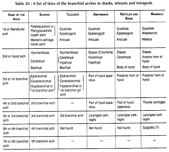
The visceral bars become united with one another to form the visceral arches. There are four to nine visceral arches in vertebrates. Of the visceral arches, the first or mandibular arch and the second or hyoid arch participate in skull-formation while the rest are known the branchial arches (Fig. 4.3B).
A list of derivatives of branchial arches in sharks, teleosts and tetrapods is given in Table 20.
In all vertebrates except the agnathans, the mandibular arch becomes modified into jaws. The dorsal elements or the palatoquadrates or pterygoquadrate cartilages become the upper jaw while the ventral pair (Meckel’s cartilages) form the lower jaw. The posterior end of the pterygoquadrate, the quadrate provides an articulation for the lower jaw.
The hyoid bar also becomes divided into two parts—a dorsal part, called hyomandibula, and a ventral part, designated as hyoid cornu. The hyoid cornu is divisible into epihyal, ceratohyal and hypohyal. The tongue is supported by a median basihyal which is attached with ceratohyal.
Jaw Suspension:
Jaws are the characteristic feature of the gnathostomes which are used for holding the prey, and chewing the food materials. In the course of vertebrate evolution, the appearance of jaws has profoundly changed the mode of living.
Before the appearance of jaws, the animals like protochordates are the ciliary feeders. The cilia of the buccal funnel or in other parts of the body play a major role in food collection. In agnathans the mouth is suctorial. The ostracoderms collected their food, mainly algae or other organisms by scraping the rock surfaces.
Origin:
The first visceral arch (mandibular arch) is modified into jaws and embraces it firmly to the chondrocranium. The upper part of the mandibular arch (epibranchial) becomes palatoquadrate. The lower part of mandibular arch (ceratobranchial) forms the lower jaw, also called Meckel’s cartilage.
The second visceral arch or hyoid arch is connected with the chondrocranium and acts as supporting structure. The upper part of second visceral arch is called hyomandibula. Next succeeding arches are called branchial arches and are connected to the respiration, not associated with the formation of jaws.
Definition:
The mechanism by which the upper jaw (palatoquadrate) and lower jaw (Meckel’s cartilage) are suspended from the neurocranium, is called jaw suspension.
Types of Jaw Suspension:
Goodrich (1930, ’58), Walter and Sayles (1949), and Weichert and Presch (1975) have mentioned 4 types of jaw suspension.
These are:
(i) Amphistylic (primitive elasmobranches),
(ii) Hyostylic (Sharks and sturgeons),
(iii) Autostylic (tetrapods other than mammals) and
(iv) Craniostylic (mammals).
Hyman (1942) has mentioned 5 types of jaw suspension.
They are as:
(i) Amphistylic (primitive sharks),
(ii) Hyostylic (most elasmobranchs),
(iii) Autostylic (most vertebrates),
(iv) Holostylic (holocephali), and
(v) Methylostylic (teleostomes).
Young (1981) has also mentioned 5 types but in different names in some groups. Kardong (2002) refers to 6 types of which pataeostylic is referred to agnathans where none of the arches attach themselves directly to the skull.
The splanchnocranium is attached with the neurocranium to form a full-fledged skull. Five principal types of such attachment are encountered in vertebrates (Fig. 4.3C-F).
They are:
Autodiaslylic, Amphistylic, Hyostylic, Autostylic and Craniostylic. In the first three types of jaw suspension the pterygoquadrate, the hyomandibular or both are involved while in the fourth variety attachment is done by investing bones.
Autodiastylic:
It is a form of jaw suspension in which the upper jaw (palatoquadrate) is suspended from two articulations with the cranium. The suspended structures are ligaments which hang both at its front as well as hind end of the cranium. The hyoid arch remains an almost typical branchial arch, neither modified, nor to support the jaw. This is the most primitive type of jaw suspension, found in acanthodians.
In some sharks such as Chlamydoselachus (frilled shark), Hexanchus (six-gilled shark), Notorhynchus (broad nose seven gill shark), the squaloids (dog fish shark), pristiophoroids (saw sharks) and squatina (angle shark) the orbital process is on the palatoquadrate and an attachment to the orbit is seen. This type of a few suspension is referred to as Orbitostylic by Maisey (1980).
Amphistylic condition (both pillar):
This type of jaw suspension is found in a few primitive elasmobranchs (like Heptranchias). In this type of jaw suspension both the pterygoquadrate and hyomandibular make direct articulation with the neurocranium (Fig. 4.3C).
The pterygoquadrate articulates at two points:
(i) A basal process and
(ii) An otic process.
The hyomandibular also articulates with the auditory capsule.
Hyostylic condition (hyoid pillar):
This type of jaw suspension is found in most modern elasmobranchs and all bony fishes. In this type of jaw suspension the hyomandibular alone acts as the suspensorium of the jaw (Fig. 4.3D).
In bony fishes (like Amia, Lepisosteus and others) the typical hyostylic condition is modified where a quadrate develops from the posterior portion of the pterygoquadrate which articulates with an articular bone. The articular is developed from the Meckel’s cartilage.
Remark:
Hyman (1942) refers to the methylostylic type of jaw suspension in case of teleostomes in which palatoquadrate is suspended mainly from the otic capsule by way of hyoid derivatives.
Autostylic condition (self-pillar):
This type of jaw suspension is found in dipnoans, amphibians, reptiles and birds where the upper jaw is articulated immovably with the neurocranium without the intervention of the hyomandibular.
The articulation is done by the quadrate of the upper jaw and the articular of the lower jaw (Fig. 4.3E). In holocephalians and lungfishes the typical autostylic condition is modified where the entire upper jaw becomes completely fused with the neurocranium.
Autostylic is divided into the following subtype.
Holostylic:
In Holocephali (Chimaeras) the upper jaw (palatoquadrate) is firmly fused to the neurocranium (brain-case) and lower jaw is suspended from it. Hyoid arch becomes complete and remains free behind. In some lizards, snakes and birds the quadrate is loosely attached.
Craniostylic condition:
This type of jaw attachment is found in mammals. This is a modified type of autostylic condition. As the articular and quadrate become transformed into malleus and incus (ear ossicles) respectively, the articulation of the jaw is done by two pairs of investing bones—the squamosals of the upper jaw and the dentaries of the lower jaw (Fig. 4.3F).
Remark:
Hyman (1942) refers to that the jaw suspension of mammals is undoubtedly derived from the autostylic type.
4. Origin of Vertebrates:
Since the inception of evolutionary concept that living organisms trans-mutated through the ages, the question of searching the ancestry of vertebrates has become a central problem in Biology. Extensive work has been done on this particular problem. The greatest difficulty confronting the workers on this line is the lack of good fossil records of their earlier representatives.
Many non-chordates have been brought to the forefront in unraveling the vertebrate evolution. It will not be an exaggeration to point out that almost all the non- chordate phyla have been suggested that hold the key of vertebrate evolution.
But many workers do not accept the idea of derivation of vertebrates directly from any specialised adult non-chordate. The dynamic larval stage of some non-chordate forms are claimed to be the possible progenitor of early chordates which have evolved further to give rise to higher forms.
A good many views are extant on the origin of vertebrates. Some of the views are old and they have been mentioned as pieces of historical importance. Since long time it was regarded that the invertebrate chordates, originating from some non-chordate source, have given the origin of the vertebrates.
The logical evolutionary trend is as follows:
Biological Organisation:
The chordates constitute a very large group which includes the invertebrate chordates and vertebrates. They possess some identifying features. These features are again repeated to draw parallelism between the vertebrates and the non-chordates in the phylogenetic discussion of the vertebrates.
These features are:
a. Body is bilaterally symmetrical.
b. A dorsal tubular nerve cord is present. The anterior part becomes specialised into a brain in the vertebrates.
c. Notochord is present at least in some stage of life-history.
d. The pharynx is perforated by gill-slits.
e. The coelom is enterocoelous in origin.
f. Cephalization is well-marked.
g. Metamerism is present.
h. A pulsating organ or heart is present in the ventral side of the body.
5. Ancestry of Vertebrates:
Similarities existing between some non-chordates and the chordates have led to the enunciation of several theories on the origin of vertebrates. All these theories postulate that the vertebrates originated either directly from some non-chordates or through the intervention of some invertebrate chordates.
Coelenterate theory:
Masterman (1897) has advocated that the bilateral symmetry and the triploblastic condition of the earlier stages of Branchiostoma have been derived from some round coelenterates. Such an idea has yielded no solution to the problem.
The coelenterates stand at a very lower level of metazoan grade of organisation and the attempt to advocate the emergence of the chordates directly from such a slowly organised group seems to be unjustified.
Nemertean worm theory:
The idea of the origin of the chordates from the nemertean worm was advanced by Hubrecht in 1887. He put forward the following arguments in support of his contention.
These are:
a. The long proboscis with its sheath in nemertean worm can be regarded as the primal source of the notochord.
b. The lateral blood vessels of the nemertean worm have shifted their positions to become the dorsal and ventral vessels of the chordates.
c. In nemertean worm, the nervous system forms eight longitudinal cords. Hubrecht and Kofoid have suggested the origin of a dorsal nerve tube at the expense of the lateral and ventral nerve cords.
d. Few apertures in the nemertean worm are compared with the gill-slits.
e. The mode of development of the photosensitive cells is similar.
Remarks:
All the points of homology cannot be proved by scientific studies. Because of the lack of evidences and of the existence of wide structural diversities, the nemertean worm concept on the origin of chordate cannot be approved.
Phoronis theory:
Masterman (1897) also tried to relate the phoronids with the chordates on a phylogenetic ground. The theory was based on the similarities existing between the Actinotrocha larva of Phoronis and the Tornaria larva. The most important evidence comes from the disposition of coelom in the two larvae.
A pair of gastric diverticula opening into the anterior end of the stomach is homologized by Masterman with the notochord. A septum separating the mesocoel and metacoel is present in Phoronis and Balanoglossus. The tentacular crown of Phoronis resembles the tentaculated arms of Cephalodiscus. The proboscis pore of Balanoglossus is comparable to the water pore of Phoronis.
Remarks:
All the above observations of Masterman have not been corroborated by anatomical evidences. The similarity on the disposition of the coelom can be interpreted as due to the consequence of common remote ancestry of all the Deuterostomous phyla. So the question of holding the ancestral position of the chordates by Phoronis cannot be justified.
Annelid theory:
The concept of the annelid ancestry of the chordates was mainly sponsored by Dohrn (1875), Semper (1876) and Minot (1897). They hold the view that the primitive chordates have been evolved from the annelids, typified by the earthworm and some marine worms. The vertebrates have evolved directly from these primitive chordates.
The theory was based on the following grounds:
a. Both the groups have bilaterally symmetrical and metameric ally segmented body.
b. The parapodia of annelids are comparable with the paired appendages of the vertebrates.
c. Well-developed coelom is present in both the groups.
d. Presence of nerve cord, although differing in disposition.
e. Presence of segmental excretory organs.
f. Presence of longitudinal blood vessels and red-coloured blood.
g. As the disposition of the nerve cord and the direction of the blood-flow through the blood vessels in annelids are diametrically opposite to that of the chordates, the workers advanced the idea that if the dorsal and ventral positions of the annelid are reversed, the worm will assume the shape of a chordate (Fig. 4.4).
But the annelid origin of chordates is not convincing because, fundamentally a chordate cannot be compared with an annelid.
i. The nature and the disposition of the nerve cord in annelids are quite different. In annelids the nerve cord is a solid structure and occupies a mid- ventral position. But in chordates the nerve cord is a mid-dorsally placed hollow tube.
ii. The nature of segmentation is also different. The segmentation in annelids shows complete division of the body into rings, whereas in chordates only the myotomal region is segmented.
iii. In the circulatory system of annelid, the direction of blood-flow through the longitudinal blood vessels is not similar to that in chordates and the haemoglobin remains dissolved in the plasma.
iv. In the chordates, the haemoglobin is contained by the R.B.C.
v. The parapodia of annelids are in no way homologous to the paired appendages of the vertebrates.
vi. The presence of notochord in chordates has no parallel in annelids, although attempts have been made to homologies the ‘Faserstrang’ (a bundle of fibres extending along the nerve cord) with the notochord.
vii. The developmental history of the annelids also differs in a number of features. The segmentation of annelids follows a spiral fashion, while in chordates the cleavage plane is radial or irregular.
viii. The mode of formation of coelom is different. In annelids the coelom is schizocoelic in origin, while in the chordates it is enterocoelic.
Remarks:
Considering all these contrasting features, the phylogenetic relationship between the annelids and the chordates cannot be established. All the alleged points of homology are mostly speculative and have no solid basis to establish the relationship.
Insect theory:
Hilaire (1818) propounded the idea that the vertebrates might have been derived directly from the insects. He put forward the following evidences in support of his idea.
These are:
a. The segmented body rings of insect are the precursors of the vertebral column.
b. The legs of the insects have in course of time formed the ribs of the vertebrates.
c. The tergum of insect can be compared with the dorsal plate of primitive fishes.
d. Cephalisation is comparable.
e. Hilaire put forward the notion that if an insect be made completely overturned, it will portray the entire vertebrate organisation.
Remarks:
This particular concept is not acceptable, because the points of resemblances are mostly imaginary. The insect theory gets a very little reorganization. Insects constitute a very specialised group of non-chordates and the emergence of vertebrates from such well-formed organisms seems to be unjustified. The whole of the developmental events of vertebrates differs greatly from that of insects.
Arachnid theory:
Attempts have been made to prove the process of descent of the vertebrates from some primitive arachnids. The theory was proposed by Patten (1912) and Gaskell (1908). They have taken the ostracoderms as the typical representative of the primitive vertebrates and have set aside Branchiostoma and other invertebrate chordates from the picture.
Limulus is regarded as a primitive member of the Arachnida. It shows structural relationships with Eurypterids (heavily armoured fossil arachnids of the Cambrian to Silurian periods) and the Eurypterids, in turn, resemble the Cephalaspids (ostracoderms) of the Devonian period.
The probable line of ancestry is as follows:
Limulus à Eurypterids à Ostracoderms
Patten wanted to show striking similarities between the heart and the arterial system of Limulus and the vertebrates. The fused cephalothoracic ganglionic mass of arachnid is comparable to the brain and cranial nerves of vertebrates. The ‘endocranium’ (sternum which protects the central ganglionic nerve complex) of Limulus resembles the primitive vertebrate cranium.
Special sense organs, specially the eyes, are comparable. Gaskell has carried the theory to the extreme by assuming that the original digestive system of arachnid has been converted into the nervous system of the vertebrates and the digestive system has been formed anew. The eurypterids and the ostracoderms have many superficial similarities, particularly in the general arrangement of the plates and head shield.
Remarks:
In spite of all efforts to execute the idea of the origin of the vertebrates from the arachnids, Patten and Gaskell have failed to advance any solution to the problem though the theory is supported by a large body of evidences.
Echinoderm theory:
Of all the theories discussed so far, the Echinoderm theory on the origin of chordates needs consideration. Many investigators have put forward the following evidences from various considerations to establish the view that the chordates have originated directly from the echinoderms:
Anatomical evidences:
The most important anatomical similarity lies in the presence of mesodermal skeleton in both the echinoderms and chordates. The nervous system in hemichordates resembles very closely that of the echinoderms. In both the cases the nervous layer is present at the basal part of the epidermis.
Embryological evidences:
The Tornaria larva of Balanoglossus and the larvae of Echinodermata exhibit close similarity (Fig. 4.5). Because of the resemblances, Johannes Muller and Bateson suggested that the Tornaria larva and the Dipleurula larva have evolved from a common ancestral source.
The common features are:
a. The body is minute and transparent.
b. Both have a dorsal pore.
c. Almost identical twisted external ciliated bands are present.
d. Formation and disposition of the coelom are similar.
e. Both are free-swimming forms.
f. Body is bilaterally symmetrical.
But the presence of apical plate with eye spots in Tornaria larva raises doubt. The probable line of ancestry is suggested but further evidences are required to establish the fact. To put more emphasis on the relationship between the echinoderms and the chordates, Garstang and De Beer have added more weight on the ancestry of the chordates from the echinoderm larva. They proposed the Neotenous Larva Theory.
They advocated the concept that the persistence of the echinoderm larva and its attainment of sexual maturity would have led to a neotenous larval phase. This particular larval form would have supplied all the raw materials for chordate evolution.
Garstang (1894) also imagined that if the ciliated bands together with the underlying nervous tissue of Auricularia larva become accentuated to form ridges leaving a groove between them and if the lips of such grooves subsequently become fused to form a tube, a structure would be produced resembling the chordate nervous system.
At last, in brief it is said that beginning with an echinoderm larva (auricularia larva) Garstang proposed a series of evolutionary stages that involved paedomorphosis and production of chordates (Fig. 4.6).
However, some more additional supports are necessary—particularly from the palaeontological side—to establish the line of ancestry. But the chances of finding conclusive fossil records of such soft-bodied larvae are very remote.
Palaeontological evidences:
The most primitive echinoderms recorded in the geological history are that of Cambrian and Ordovician Carpoid echinoderms. Torsten Gislen (1930) assumed that the Carpoid echinoderms might have evolved from tornaria-like creatures which have begun to settle down to lead a sedentary life. The water vascular system might have developed out of the ciliated grooves of the tornaria-like forms.
It has also been claimed that, in the lower Silurian, one of the Carpoid echinoderms had the calyx perforated by a series of 16 small apertures. These apertures are compared with the gill-slits of Branchiostoma. The carpoid echinoderms constitute a very divergent group and some forms also show a superficial similarity with some pteraspid ostracoderms.
R. P. S. Jeffries published a book in 1986 — The Ancestry of Vertebrates. In this book he proposes that vertebrates and other living deuterostomes each originated from a palaeozoic fossil group — the calcichordates, which T. Gislen (1930) considered as echinoderms but R. P. S. Jeffries contends that they are chordates.
Biochemical evidences:
The phylogenetic relationship between the echinoderms and the chordates has been shown by biochemical studies also. Most of the non-chordates conduct energy transfer with arginine phosphate, but ophiuroids, cephalochordates, ascidians and vertebrates use creatine phosphate. The hemichordates and echinoids use both arginine phosphate as well as creatine phosphate.
J. Needham (1932) has shown the presence of phosphogen as the phosphorus-carrier in the echinoderms and chordates. Wilhelmi (1942) has shown the affinity between the two groups by serological tests. All these isolated biochemical studies put weight on the concept of derivation of the chordates from the echinoderms.
Ancestry of Chordates:
None of the theories except the echinoderm theory has received attention, no matter how skilfully the theories have been presented. Neither annelids nor arthropods can be regarded as the ancestral form of the chordates. An effort to derive a highly specialised group from another highly modified one can never be logical.
The natural tendency uppermost today is the recognition of a true relationship between echinoderms and chordates, because the ancestral garment, according to many workers, fits well upon the group.
Attempts have been made to solve the problem from the following angles:
a. Consideration of relationship in larval organisation.
b. Evidences deriving from the morphological studies.
c. Supports from the biochemical experimentations.
d. By comparing fossil echinoderms with the fossil chordates.
By setting aside the controversies on this issue, it is accepted by many that the dynamic larval forms of echinoderms hold the key of chordate ancestry. Many workers incline to follow Garstang’s view that the free-swimming auricularian larva by paedomorphosis has given origin to the chordates.
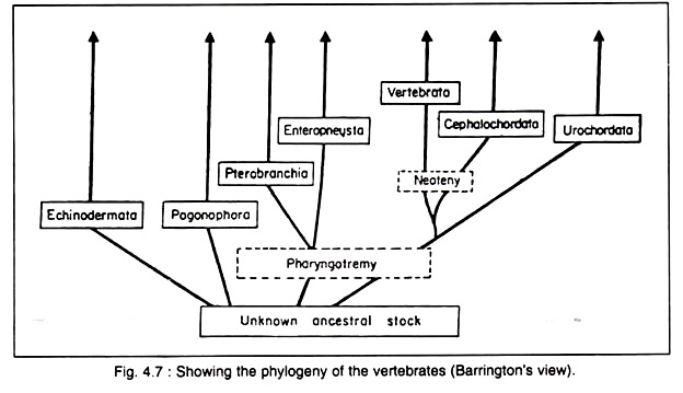
But no convincing evidence is ready at hand to explain the process of such transformation. Barrington (1965) and many other workers do not accept the emergence of the chordates directly from the echinoderms. According to Barrington the members of the Deuterostomia (Echinodermata, Pogonophora, Invertebrate chordates and Vertebrata) have originated from a common ancestor (Fig. 4.7) having:
i. Bilaterally symmetrical body,
ii. Sessile or semi-sessile habit,
iii. Tripartite body and coelom,
iv. Coelom with coelomopores,
v. External food collection and
vi. Ciliated larval form.
The echinoderms do not hold the key of chordate origin but represent a sideline evolution from the same ancestral source.
Origin of the Vertebrates:
Claims have been made by many that vertebrates have either been evolved from some non-chordate source or through some Invertebrate chordates. But the idea of derivation of the vertebrates from some invertebrate chordates needs thorough reconsideration in the light of present investigations.
Branchiostoma ancestry:
The theory of the origin of vertebrates from Branchiostoma appears to be convincing, because it fulfils the theoretical definition of a typical chordate organisation. Special emphasis has been put on the presence of notochord, dorsal tubular nerve cord and the pharyngeal gill-slits.
For this reason it was believed that Branchiostoma has channelized the line of vertebrate evolution. It has a fish-like appearance and gives a fairly typical picture of an early stage in the vertebrate evolution. Branchiostoma exhibits many structural similarities with the Ammocoetes larva of Lamprey.
Both of them possess the following common characteristics:
(a) ‘V’-shaped myotomes.
(b) Oral hood.
(c) Continuous dorsal fin.
(d) Velum and velar tentacles.
(e) Endostyle.
Due to the structural resemblances with the Ammocoetes larva, it is suspected by many that the grade just above Branchiostoma in the evolution of vertebrate is well-represented by Ammocoetes larva. But difficulties stand in assuming Branchiostoma as the true ‘stem-form’ in the evolution of vertebrates.
Though it is undeniable that it has many primitive chordate characteristics, yet it may, at best, be regarded as the primitive chordate. A thorough survey of the anatomical organisation reveals the existence of many specialised as well as degenerated features. Such highly specialized and degenerated form cannot possibly react to the evolutionary dynamicity of the vertebrates.
The structural organisation throws much light on the probable ancestral vertebrate form. Branchiostoma stands nearest to the true ancestral vertebrates but its sedentary habit offers the strongest objection in assuming the biological changes of the vertebrates from sessile animal like Branchiostoma.
Gutmann (1981) proposes that the earliest chordates were amphioxus-like animal and other groups of animals like sessile urochordates, vertebrates and burrowing hemi-chordates are developed from this group. Benthic echinoderms originate eventually from the burrowing hemichordates (Fig. 4.8).
Balanoglossus ancestry:
Balanoglossus is regarded as the ancestor of the vertebrates. The Tornaria larva shows marked resemblance with the echinoderm larvae. Wilder regarded the evolutionary succession of the hypothetical echinoderm ancestor into Balanoglossus and Balanoglossus to the vertebrates.
But the fallacy lies in the fact that Balanoglossus itself does not fulfil the standard criterion of a standard chordate. The simpler organisation of Balanoglossus cannot be denied but its degeneration is questionable. The so-called notochord (anterior extension of the dorsal wall of the pharynx) is a very doubtful structure. All the recent workers on this line do not accept this structure as the notochord.
The nervous system in Balanoglossus is more of non-chordate type than that of the chordate. So the only important chordate characteristic that remains is the presence of pharyngeal gill-slits. The similarities of Balanoglossus with the vertebrates are due to remote phylogenetic relationship. The hemichordates as a group evolved from unknown sessile or semi-sessile ancestors having bilaterally symmetrical body with tripartite body and coelom.
The hemichordates have remained closer than other non-chordates, especially the echinoderms and pogonophores. The hemichordates have developed pharyngotremy—an important feature for chordate organisation. Evolution of pharyngotremy is associated with the ciliary mode of feeding.
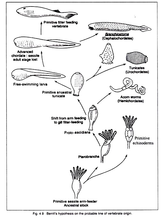
Urochordate ancestry. N. J. Berrill in his book “Origin of Vertebrates” (1955) attempts to find a line of approach on the problem of vertebrate evolution. Urochordates have long been removed from the picture of the ancestry of vertebrates. But Berrill has tried to bring the urochordates to the forefront of vertebrate evolution.
According to him, the question of degeneration is nothing anew in lower chordates. The sedentary habit of the urochordates shows close conformity with other sedentary invertebrate chordates. Berrill wants to impress on the fact that the free-swimming tadpole larva of urochordates has come from the sessile life of the vertebrates.
This idea forces us to abandon the long accepted notion that from the free-swimming larvae sessile condition in the adult came into existence.
Berrill thinks that the free-swimming tadpole larva—through developmental paedomorphosis (i.e., effectively exhausting the potencies of re-differentiation of cells through certain unknown factors)—has avoided the sedentary condition and maintains a perennial free- living habit.
He adds no importance to Branchiostoma as the direct ancestor of vertebrate and advocates that the immobile habit cannot possibly give rise to mobile vertebrates. Balanoglossus cannot hold the key of vertebrate line of descent. So Berrill is driven to the inevitable conclusion that the dynamic tadpole larva of urochordates is the actual source from which vertebrates have been evolved in time and space.
Berrill has put forward the following arguments to support his interpretation. According to him the tadpole larva in urochordate has been evolved within the group to fulfill certain specific ascidian needs. In course of development some tadpole larvae became neotenous and ceased to metamorphose into adults.
They started reproducing sexually as free-swimming organisms. This phenomenon had taken place in the oceanic surface water. During the earlier part of the pelagic evolutionary phase some members used to exploit the river mouth. The effective locomotory power has enabled the tadpole larvae to make an excursion into the far extent in the river system.
At first, such excursion into the river system was intended solely for feeding and they used to return to the ocean for breeding. Branchiostoma, as we see today, is one of the relics of such transitional phase which rediscovered some advantages of oceanic life and became re-adapted to it.
Within the river system, such tadpole larvae directly evolved into relatively simple unarmed ostracoderms. Such ostracoderms represent the vertebrate prototype which has given origin to the specialised ostracoderms and, subsequently, to the true fishes.
The probable line of evolution of Vertebrates as visualised by Berrill is shown in Fig. 4.9. The concept of Berrill regarding the origin of Vertebrates from the tadpole larva of Ascidia is not accepted at present. Nowadays, there is a general trend to accept Barrington’s view on the origin of vertebrates.
Probable Ancestry of Vertebrates:
Because of scanty information, the discussion on the origin of Vertebrates becomes a complicated and debatable issue. Baldwin (1963), Ennor and Morrisson (1958) and many others have shown phylogenetic relationship on the distribution of phosphagens—the phosphate carriers which participate in energy- production in muscular activities.
They have established that phosphagen—in the form of phosphocreatine—is present in Amphioxus and Vertebrates whereas phosphoarginine is present in majority of the non-chordates and urochordates. Both these compounds are detected in echinoderms and hemichordates.
From the above evidences it is advocated that the non-chordates and vertebrates are related to echinoderms and hemichordates, but it throws urochordates in an anomalous situation. Above observation justifies the notion that the urochordates are situated far from the main chordate line.
But the presence of phosphocreatine in the muscles of an ascidian species, Pyura, added complication to the issue. The phylogenetic value of the existence of phosphagens in evaluating the relationship between different groups is doubtful.
Presence of common metabolic pathways in different groups may be interpreted as the instance of independent evolution of similar biochemical adaptations. But the distribution of phosphagens fits well with the current views regarding the relationships of the deuterostomia which is summarised below.
ADVERTISEMENTS:
The echinoderms, pogonophores, hemichordates, urochordates, cephalochordates and vertebrates belonging to a common group Deuterostomes are phylogenetically related with one another and they have evolved from common ancestral stock which had bilaterally symmetrical bodies, tripartite body and coelom, external food collection, ciliated larval form and sessile or semi-sessile habit.
The protochordates (hemichordates, urochordates and cephalochordates) form a sort of connecting link between the vertebrates and the rest of the deuterostomes. The protochordates are highly specialised forms and it is different to ascertain the straightforward sequence of stages leading to the origin of Vertebrates.
But the phylogenetic status of the hemichordates is difficult to establish. The cephalochordates and urochordates are true chordate but their relationships with each other as well as with the vertebrates are difficult to ascertain.
The deuterostomes have originated from sessile or semi-sessile ancestral form. The ancestor had bilaterally symmetrical tripartite body and coelom. They used to collect food externally by ciliary method. The echinoderms and pogonophores have departed earlier from the ancestral stock whereas the hemichordates stand closer to them.
The hemichordates developed pharyngotremy to assist respiration and feeding mechanism. In course of time and space a group evolved which had specialised pharyngeal apparatus to assist interval food collection.
The group holds the key of the origin of vertebrates, cephalochordates and urochordates. The larva of the group (possibly derived from ciliated larva) became specialised for pelagic life and, by neoteny, gave origin to free-swimming adults. These free-swimming adults gave origin to the Vertebrates and Cephalochordates (Fig. 4.8).
Modern cephalochordates failed to exploit the advantages of pelagic life and the adults became semi-sessile. The urochordates retained the sessile habit in adults while the larvae are dynamic for habitat selection. The hemichordates are lowly organised and primitive invertebrate member of the Phylum Chordata.
They are separated from the urochordates and cephalochordates by a wide gap. The inclusion of urochordates and cephalochordates under the Phylum Chordata is quite obvious.
But the sessile adult of some protochordates put difficulty to the problem. Garstang (1928) developed the idea that the chordates began with sessile or semi-sessile forms and the cephalochordates and vertebrates evolved from such forms with the loss of sessile adult and retention of free-swimming larval stage.
If their concept is taken to be true, then the life-cycles of the protochordates have to be modified in a way that sexual maturity is achieved in larval phase (Neoteny). The concept of neoteny as a factor of evolution is quite convincing.
Time of origin:
The chordates constitute very ancient forms and the tangible fragmentary remains of the invertebrate chordates have been recorded in the Cambrian strata and that of ostracoderms in the middle Ordovician time.
But the relics discovered in the strata are of those creatures who had already travelled a long evolutionary way. It can be said that the time of origin was not later than the beginning of the Cambrian, if not long before.
Place of origin:
The place of origin is not clear. There are evidences that the vertebrates first appeared in the sea and migration into the river system took place during the early phase of vertebrate evolution. From the discovery of the earliest chordates in the sea, it is believed that the marine environment is the site of the origin of chordates.


